
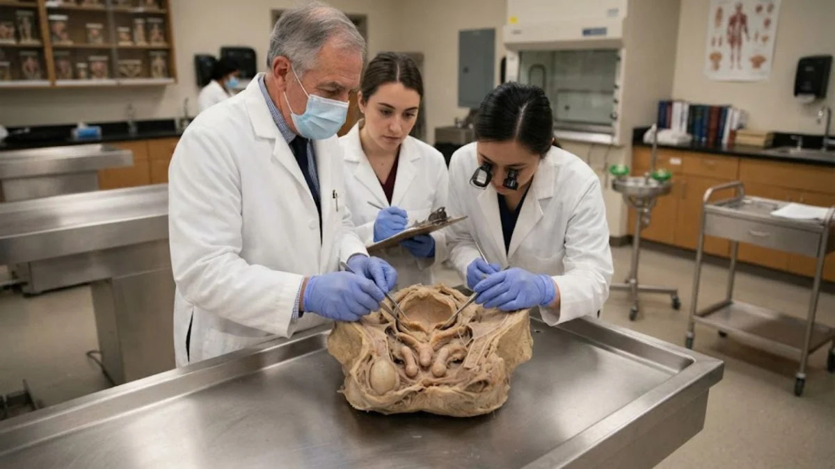
"This remarkably concealed case of triphallia underscores that the true prevalence of supernumerary penises may be significantly underestimated, as many instances likely go undetected due to their lack of external manifestation and absence of symptoms."
The discovery of a donated pelvis at the University of Birmingham Medical School revealed an extraordinary anatomical anomaly: not one, but three penile structures stacked one behind the other within the scrotum of a white man in his late 70s. This case, meticulously documented in the Journal of Medical Case Reports, represents only the second known instance of triphallia in humans. While the sheer number of penises initially garnered significant attention, the true significance of this finding lies in its profound implications for our understanding of anatomical variations and the potential for widespread underdiagnosis of polyphallia, particularly in cases where external signs are absent. The authors suggest that the documented prevalence, derived from cases that are externally visible or symptomatic, may not reflect the full picture, raising critical questions for clinical practice and anatomical classification.
The donor, a man of approximately six feet in height, was subjected to a routine teaching dissection. Under the established protocols of the University of Birmingham Medical School, specific identifying information and detailed medical history of the donor were withheld from the research team, ensuring donor anonymity. The dissection proceeded by dividing the pelvis along the midline, a standard procedure for anatomical study. It was during this meticulous process, as the pubic symphysis, penis, and scrotum were transected in the midsagittal plane, that the researchers encountered the unexpected internal structures.
Aligned in a front-to-back orientation, a configuration described by anatomists as sagittal stacking, were three distinct penis-like formations. The most dorsal, or uppermost, structure was the only one externally visible, appearing as a typical penis. Immediately beneath this was a second, fully formed penile structure. This secondary penis possessed its own corpora cavernosa (the erectile tissues), corpus spongiosum (the tissue surrounding the urethra), a complete urethra, and a glans. The third, and deepest, structure was also partially developed, featuring corpora cavernosa and a glans, but notably lacked a corpus spongiosum and a urethra.
The clinical significance of this case was further illuminated by the functional investigation of the urethral pathway. Upon insertion of a probe into the external urethral opening, it failed to advance. Subsequent dissection revealed the reason for this obstruction: a single, continuous urethra originated from the bladder outlet and traversed through the secondary penis before merging into the primary penis. This meandering course was characterized by its singular, unbranched nature.
A particularly noteworthy aspect of this case was the absence of associated anomalies that frequently accompany penile duplication. Typically, such conditions are linked with a spectrum of congenital defects, including accessory kidneys, ureters, or bladders; duplication of the lower gastrointestinal tract; a bifid scrotum; an imperforate anus; or abdominal wall defects. In contrast, this particular instance of triphallia presented without any of these commonly associated malformations, making it an outlier compared to many externally visible cases.
The prevailing understanding of the incidence of supernumerary penile formation, or polyphallia, has historically been based on figures suggesting it affects approximately 1 in 5 to 6 million live births. This estimate is largely derived from the report of the first documented case of triphallia, which involved a three-month-old infant in Duhok, Iraq. A comprehensive review of literature by the Birmingham team, encompassing 168 papers published between 1606 and 2023, identified 112 cases of complete diphallia (two penises), 50 cases of pseudodiphallia (partial duplication), and, prior to this new finding, only one confirmed case of triphallia. This classification adhered to a framework established in the 1950s, with five papers deemed to lack sufficient detail for precise categorization.
However, the current findings challenge these statistical assumptions. The authors propose that the recorded prevalence is heavily skewed towards cases that manifest externally or present with clinical symptoms. In this particular instance, the three penile structures were entirely concealed, meaning the donor’s external anatomy would have appeared entirely normal during any routine physical examination. Furthermore, he had experienced no discernible symptoms that would have prompted further investigation into this anomaly. The authors’ conclusion, therefore, is that "the prevalence of polyphallia may be greater than expected."
The logic underpinning this assertion is straightforward: the published data primarily reflects those cases that are readily apparent at birth or later in life through urinary or sexual complications. In such symptomatic cases, surgical intervention, typically the removal of the less functional or smaller extra penis, is a common course of action. Reports of diphallia cases with internal accessory penises are rare, with only six documented instances. Concealed cases, like the one under discussion, which present without any overt signs or symptoms, leave no trace in the medical literature, thus contributing to an artificially low reported incidence.
A critical detail that circulated widely following the initial dissemination of this case was the assertion that the man lived his entire life unaware of his condition. The authors of the case report, however, exercise greater caution and nuance in their conclusions. They explicitly state that they cannot definitively confirm that the anomaly went unnoticed throughout the donor’s life. Their reasoning is grounded in the documented history of inguinal hernia repair. Such a surgical procedure in the inguinal region would have involved direct visual inspection of the area by a medical professional, who might have observed or at least encountered the unusual anatomy during the operation.
Similarly, the authors are measured in their assessment of potential consequences. In cases involving blind-ending or duplicated urethras, the risk of urine stagnation and subsequent infection is elevated, a known clinical concern. However, in this specific case, the absence of a blind-ending urethra led the researchers to judge that the risk of infection was unlikely to be significantly heightened. They offer speculation that the concealed structures might have caused discomfort or pain during sexual intercourse if they became erect, but emphasize that this remains an unsubstantiated hypothesis.
The practical implications of this discovery extend to common clinical procedures. The authors highlight that the tortuous path of the single, albeit shared, urethra would have posed a significant challenge for the passage of a urinary catheter. A clinician attempting catheterization on a patient with outwardly normal anatomy, encountering unexplained resistance, would have no immediate reason to suspect an underlying internal duplication of this magnitude. The potential for iatrogenic injury in such a scenario is considerable, especially when the patient is unable to provide a warning about an unknown anatomical variation. Therefore, the authors strongly advocate for increased awareness of such possibilities among healthcare professionals. This awareness is crucial for the safe and effective performance of procedures such as catheter insertion, urological imaging, and surgical interventions. Moreover, it is a factor that should be considered when evaluating patients presenting with unexplained urological symptoms.
Beyond the immediate clinical considerations, this case also exposes a definitional and classificatory challenge within medical science. There is a recognized lack of a comprehensive and clinically practical classification system for supernumerary penises. The widely adopted Schneider framework, for instance, does not adequately account for the diverse phenotypes of these anomalies. While a more recent system proposed by Kendrick and Kimble offers a more detailed categorization, its increased complexity can limit its immediate utility at the bedside. The authors of the triphallia report call for the development of a simpler, more clinically applicable classification system to better guide diagnosis and management.
It is imperative to reiterate that this case stems from a single cadaveric study. Consequently, it cannot provide quantitative data on the actual prevalence of polyphallia. However, its singular existence demonstrates that at least one case of a significantly complex anatomical variation occurred, one that evaded detection during all living examinations and remained unrecorded in clinical histories. This finding, while unsettling, serves as a powerful reminder of the vast and often unacknowledged spectrum of human anatomical diversity and the limitations of our current diagnostic capabilities in identifying all such variations.
