
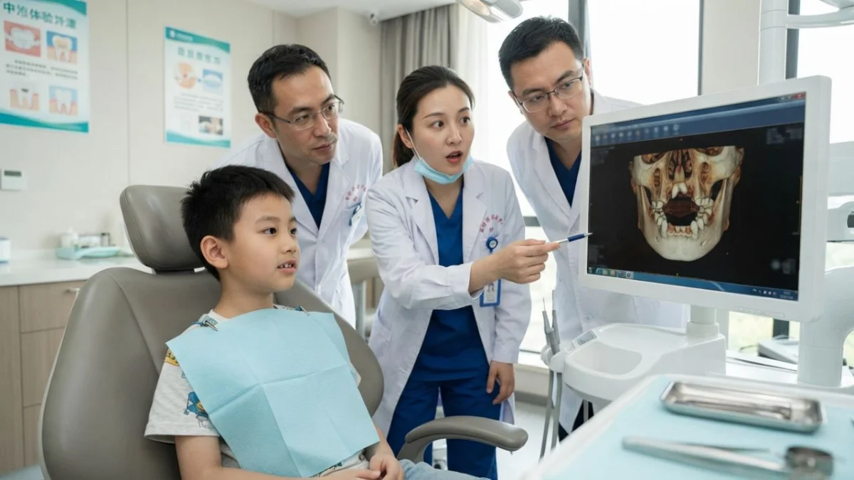
A rare case of an inverted supernumerary tooth, lodged upside down in a young boy’s palate and pushing towards his nasal cavity, highlights the often-overlooked dental origins of seemingly unrelated medical symptoms. This extraordinary dental anomaly, which mimicked persistent allergies, was only discovered after the child sought treatment for a chipped front tooth, underscoring the importance of interdisciplinary collaboration between dental and medical professionals.
A 9-year-old boy in China presented at a dental clinic with a chipped front tooth, a seemingly straightforward dental injury. However, his visit unexpectedly led to the diagnosis of a rare dental anomaly that had been causing him a month of discomfort: an extra tooth growing upside down, with its crown embedded in the bone of his palate and its tip directed towards his nasal cavity. This discovery provided a long-awaited explanation for his persistent stuffy nose, a symptom that had previously been misdiagnosed as allergic rhinitis and treated unsuccessfully with nasal steroids.
The unusual case, detailed in the April 7, 2026, issue of the peer-reviewed journal Frontiers in Medicine, describes how the boy had been suffering from a blocked right nostril and a dull ache in his nose for approximately one month. An initial consultation with an ear, nose, and throat specialist led to a diagnosis of allergic rhinitis, and he was prescribed mometasone furoate, a corticosteroid nasal spray. While this treatment offered some partial relief, the underlying cause remained unaddressed.
The authors of the report, affiliated with hospitals in Longyou and Jiaxing in eastern China, aptly titled their findings "An overlooked inverted supernumerary tooth." This peculiar dental intruder had been residing within his upper jaw, concealed from the view of the specialist focused on his nasal passages. The seemingly minor dental injury of a chipped tooth inadvertently provided the crucial clue to unraveling the mystery behind his prolonged nasal symptoms.
A Dental Injury Exposes the Underlying Problem
The boy’s initial dental examination was prompted by trauma to his upper front teeth, which resulted in an enamel-only fracture on one of his incisors, without any signs of pulp exposure. A visual inspection of his palate and gums revealed no swelling, redness, or discharge, suggesting a localized dental issue. It was during the detailed history-taking that the boy mentioned his persistent blocked nose and intermittent nasal pain, prompting further investigation beyond the initial dental concern.
A panoramic X-ray, a standard imaging technique in dentistry, revealed an unexpected finding: a supernumerary, or extra, tooth situated in the anterior portion of his upper jaw, slightly to the left of the midline. Critically, the X-ray indicated that this extra tooth was oriented incorrectly, with its crown pointing in an unusual direction.
While the panoramic X-ray was instrumental in identifying the presence of the supernumerary tooth, it lacked the detailed resolution required to accurately plan a surgical intervention. The flat, two-dimensional nature of the X-ray made it difficult to ascertain the tooth’s precise orientation, its depth within the bone, and its exact relationship to the hard palate, the nasal cavity, and the roots of adjacent permanent teeth. To obtain this vital information, the medical team opted for a more advanced imaging modality: cone-beam computed tomography (CBCT).
Mapping an Upside-Down Tooth in 3D with CBCT
Cone-beam computed tomography (CBCT) is a specialized dental imaging technique that generates a three-dimensional (3D) reconstruction of the teeth, jawbones, and surrounding structures. The CBCT scans provided a remarkably clear and detailed picture of the inverted supernumerary tooth, painting a complex anatomical puzzle. The images confirmed that the extra tooth was positioned upside down, with its crown facing upwards towards the nasal cavity and its root directed downwards towards the oral cavity. The crown of the tooth had already breached the outer layer of the hard palate’s bone.
Adjacent to the crown of the inverted tooth, in close proximity to the nasal lining, the CBCT scans revealed what appeared to be follicular tissue. This is the sac-like structure that surrounds a developing tooth, and its presence suggested a potential inflammatory response. The tip of the tooth’s root was located approximately 3 millimeters above the alveolar bone crest, perilously close to the root of a permanent incisor and the nasopalatine canal. This canal is a vital anatomical passage that transmits nerves and blood vessels through the anterior part of the palate, underscoring the delicate nature of the situation.
The authors emphasized the limitations of traditional flat X-rays, which often suffer from overlapping structures and a lack of fine detail. In contrast, CBCT allowed for visualization of the jaw in three distinct planes and the creation of a precise 3D model of the anomalous tooth. This detailed anatomical mapping served as an invaluable "working map" for the surgical team, enabling them to meticulously plan the extraction procedure and anticipate potential challenges.
However, the advanced imaging capabilities of CBCT come with a consideration regarding radiation exposure. The U.S. Food and Drug Administration (FDA) advises that while CBCT is a valuable diagnostic tool, radiation concerns are particularly relevant for younger patients. Therefore, dental X-ray examinations, including CBCT, should be performed judiciously, utilizing the lowest radiation dose that still yields diagnostically useful images, and only when clinically indicated.
Surgical Extraction: A Delicate Procedure Through the Mouth
The surgical approach chosen for the removal of the inverted supernumerary tooth was through the oral cavity, minimizing invasiveness and potential complications associated with nasal surgery. Under local anesthesia, surgeons made an incision along the gum line on the palatal side of the upper front teeth, carefully dissecting and elevating a flap of gum tissue to expose the underlying bone.
A piezosurgery unit, an ultrasonic bone-cutting instrument, was employed to create a precise and controlled window in the bone, granting access to the anomalous tooth. The surgical team worked with deliberate, gentle rotational movements, exercising extreme caution to avoid any upward pressure that could inadvertently drive the tooth further into the nasal cavity. This meticulous technique ensured the safe removal of the tooth in a single piece. Following the extraction, the follicular tissue was carefully curetted, and the surgical site was closed with sutures.
The postoperative recovery for the young patient was remarkably swift and successful. According to the report, the boy’s nasal blockage and associated pain completely resolved starting from the first day after the surgery. A follow-up CBCT scan performed one week post-operation confirmed the complete removal of the tooth and the integrity of the floor of the nasal cavity. At both one-month and three-month follow-up appointments, the boy remained symptom-free, his nearby permanent teeth tested normally for vitality, and his palate had healed without any visible scarring.
The Spectrum of Supernumerary Teeth: From Common to Extremely Rare
Supernumerary teeth, also known as extra teeth, are a relatively common dental anomaly, with reported prevalence rates ranging from approximately 0.3% to 3.5% in the general population. These anomalies are more frequently observed in males than in females. A comprehensive literature review published in 2010 on mesiodens, the most common type of supernumerary tooth, indicated that prevalence rates can vary significantly across different geographic populations and ethnic groups, with a consistent observation of a higher incidence in males.
Mesiodens typically appear as small, peg-shaped teeth that develop between the two upper central incisors. While often asymptomatic, they can lead to a variety of dental complications. These include delayed eruption of permanent teeth, overcrowding of teeth, displacement of adjacent teeth, and the formation of cysts. In some instances, a mesiodens can erupt in an abnormal location, occasionally even extending into the nasal cavity.
However, the case of the 9-year-old boy represents a far more complex and unusual scenario. A completely inverted supernumerary tooth that perforates the palate and extends towards the nasal cavity is considered an "extremely rare" occurrence. The authors highlighted that such cases carry a significant risk of misdiagnosis, as unilateral nasal symptoms, such as persistent blockage or pain, can easily be mistaken for common conditions like allergic rhinitis or sinusitis.
This case bears some similarity to a previously reported incident in September, where MedicalDaily covered the endoscopic removal of a tooth that had erupted inside the nose of a 45-year-old man in Nepal. The Chinese case, however, differed in the patient’s age and the method of extraction. In the current case, the anomaly was identified by a dentist in a child and surgically removed through the oral cavity.
While this single case report offers a fascinating glimpse into a rare medical phenomenon, it is important to note that it cannot definitively establish the frequency with which children’s apparent allergies are actually rooted in dental issues. The vast majority of nasal congestion in children has far more common and benign causes. Nevertheless, the authors strongly advocate for clinicians to maintain a high index of suspicion for dental etiologies in children presenting with persistent, unilateral nasal symptoms that fail to respond to conventional treatments. They further emphasize the critical need for enhanced collaboration and communication between dental professionals and ear, nose, and throat specialists to ensure comprehensive patient care and accurate diagnosis.
