
A groundbreaking surgical intervention, performed on an unborn baby still inside the womb, has successfully corrected a severe birth defect, marking a significant advancement in fetal medicine and offering a new paradigm for the treatment of complex congenital conditions. This innovative procedure bypasses the traditional post-natal challenges, potentially transforming outcomes for infants with life-threatening anomalies.
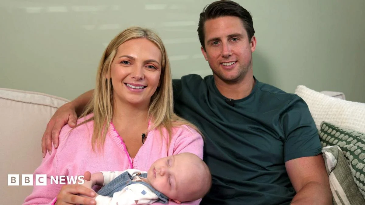
This unprecedented medical achievement centers on baby Theo, now five months old and thriving, who was diagnosed with gastroschisis – a serious birth defect where the abdominal wall fails to close, allowing the intestines to develop outside the body. His mother, Maisie Savage, underwent a pioneering in-utero surgery at 26 weeks gestation in the United States, a procedure that gently returned Theo’s exposed organs to his abdomen through keyhole surgery. This intricate intervention, part of a first-of-its-kind clinical trial, represents a monumental leap forward in pediatric and fetal surgery, offering hope where previously only arduous post-natal care and uncertain prognoses existed.
Gastroschisis, a congenital anomaly affecting approximately one in 3,000 babies in the UK each year, presents a significant medical challenge. In this condition, the abdominal muscles and skin do not form completely during fetal development, resulting in a hole, typically to the right of the umbilical cord, through which the intestines and sometimes other abdominal organs protrude. Unlike omphalocele, where organs are covered by a sac, in gastroschisis, the exposed organs float freely in the amniotic fluid. This prolonged exposure to amniotic fluid can cause damage to the delicate intestinal tissues, leading to inflammation, thickening, and shortening of the bowel, which severely impacts its function.
Traditionally, babies born with gastroschisis undergo immediate surgical repair after birth. The complexity of post-natal treatment varies significantly based on the severity of the defect. In less severe cases, often termed "simple gastroschisis," where only a portion of the small intestine is exposed, a single surgery shortly after birth may suffice to reposition the organs and close the abdominal wall. However, even these cases often necessitate weeks of intensive care, specialized nutritional feeding via a tube or intravenously (parenteral nutrition), and meticulous monitoring for complications such such as infection, intestinal obstruction, or impaired bowel function.
For cases like Theo’s, classified as "complex gastroschisis," the challenges are far more daunting. Complex gastroschisis involves additional complications such as intestinal atresia (a blockage or absence of a portion of the intestine), necrosis (tissue death), perforation, or a significantly larger amount of exposed bowel. The prognosis for these infants is considerably graver, often requiring months, sometimes up to six months, in a neonatal intensive care unit (NICU). They frequently endure multiple corrective surgeries, prolonged periods of intravenous nutritional feeding which can extend for up to two years, and in the most severe scenarios, may even require an intestinal transplant. Despite the highest standards of medical care, the mortality rate for babies with complex gastroschisis remains tragically high, with one in ten succumbing to the condition.
The rationale behind performing surgery while the baby is still in the womb is profoundly impactful. By correcting the defect before birth, surgeons aim to mitigate the damaging effects of amniotic fluid exposure on the intestines, allowing the organs to develop within the protective confines of the abdominal cavity. This early intervention seeks to prevent inflammation, preserve bowel length and function, and promote more natural growth and development of the abdominal wall, which is often underdeveloped in gastroschisis. The goal is to improve intestinal viability and function, reduce the need for extensive post-natal interventions, and ultimately enhance the baby’s long-term health outcomes.
Maisie and Josh, both dedicated teachers from London, faced an emotionally harrowing journey. The initial 20-week scan, anticipated with excitement for a first glimpse of their unborn child, quickly turned into a moment of profound shock and fear upon the discovery of Theo’s gastroschisis. Josh described the news as "incredibly scary," a sentiment shared by countless expectant parents receiving such diagnoses. Specialists at London’s Great Ormond Street Hospital (GOSH), a leading institution in pediatric care, initially prepared the couple for the arduous six months that Theo’s life would entail after birth. However, upon determining the more severe nature of Theo’s complex gastroschisis, GOSH doctors recognized the unique opportunity presented by a groundbreaking clinical trial.
This trial, spearheaded by a multidisciplinary team of fetal and pediatric surgeons at Texas Children’s Hospital in Houston, represented a beacon of hope. Led by Dr. Michael Belfort, an internationally renowned expert in fetal surgery, the team was pioneering an in-utero repair technique for complex gastroschisis. Dr. Belfort brought a wealth of experience to this endeavor, having previously innovated similar in-utero surgical procedures for babies with spina bifida, a testament to his commitment to pushing the boundaries of fetal medicine.
The decision for Maisie to travel to Texas and remain there for the remainder of her pregnancy was immense, fraught with logistical and emotional complexities. It was a choice that underscored the family’s unwavering commitment to giving Theo the best possible chance at life. Before the main surgery in Texas, a crucial preparatory step was undertaken at Leuven Hospital in Belgium. There, specialists administered a Botox injection through Maisie’s womb into Theo’s abdominal wall. This innovative application of Botox served a specific medical purpose: to temporarily relax the abdominal muscles. By inducing muscle relaxation, the procedure aimed to facilitate the easier repositioning of Theo’s organs back into his abdomen and to encourage the growth and expansion of the abdominal cavity, making the eventual repair more successful.
In November of last year, at 26 weeks gestation, Maisie underwent the highly anticipated operation in Texas. The surgical team, a confluence of expertise, meticulously performed the intricate procedure. Josh endured an agonizing wait, describing the day as feeling "like a week," consumed by uncertainty and the profound weight of the unknown. The procedure involved partially exposing Maisie’s womb to allow for optimal positioning of the uterus and the baby. The amniotic fluid, which would obstruct the surgical view, was carefully drained and replaced with carbon dioxide (CO2) gas. This gas created a clear, expanded operating space, allowing the surgeons to work with precision. Using advanced keyhole surgery techniques, Theo’s delicate organs were gently guided back inside his abdominal cavity, which was then carefully stitched closed.
The recovery for Maisie was unique and challenging. Unlike a typical C-section recovery, she was left with a significant scar, roughly twice the size of a standard C-section incision, but with the added dimension of still carrying a developing baby for another 14 weeks. Theo’s movements, particularly his kicks against the healing scar, caused her considerable pain. "It was a hard recovery," Maisie reflected, "but, it was worth it." This statement encapsulates the profound love and sacrifice inherent in such a journey.
The operation proved to be a resounding success. Theo was carried to term and born vaginally in Texas in February, a rare and positive outcome for an infant who had undergone such extensive in-utero intervention. Dr. Belfort, who also serves as obstetrician and gynecologist-in-chief at Texas Children’s Hospital, expressed immense satisfaction with the results. He highlighted the remarkable confluence of factors contributing to this success: "The mother, the baby, the surgery, the time of the surgery, the fact that we had zero complications during the fetal surgery and after the fetal surgery, just remarkable."
Theo’s story is more than just a medical marvel; it is a testament to the relentless pursuit of innovative solutions in fetal medicine. The success of this clinical trial has profound implications for the future treatment of complex gastroschisis and potentially other congenital anomalies. It underscores the growing understanding of fetal development and the increasing sophistication of surgical techniques that allow intervention before birth. This approach minimizes exposure to a hostile intrauterine environment, promotes better organ development, and significantly reduces the morbidity and mortality associated with traditional post-natal management. As Theo continues to thrive, his journey offers not only immense relief and joy to his parents but also renewed hope to countless families worldwide facing similar daunting diagnoses, paving the way for a new era in prenatal intervention and improved outcomes for the most vulnerable among us.
