
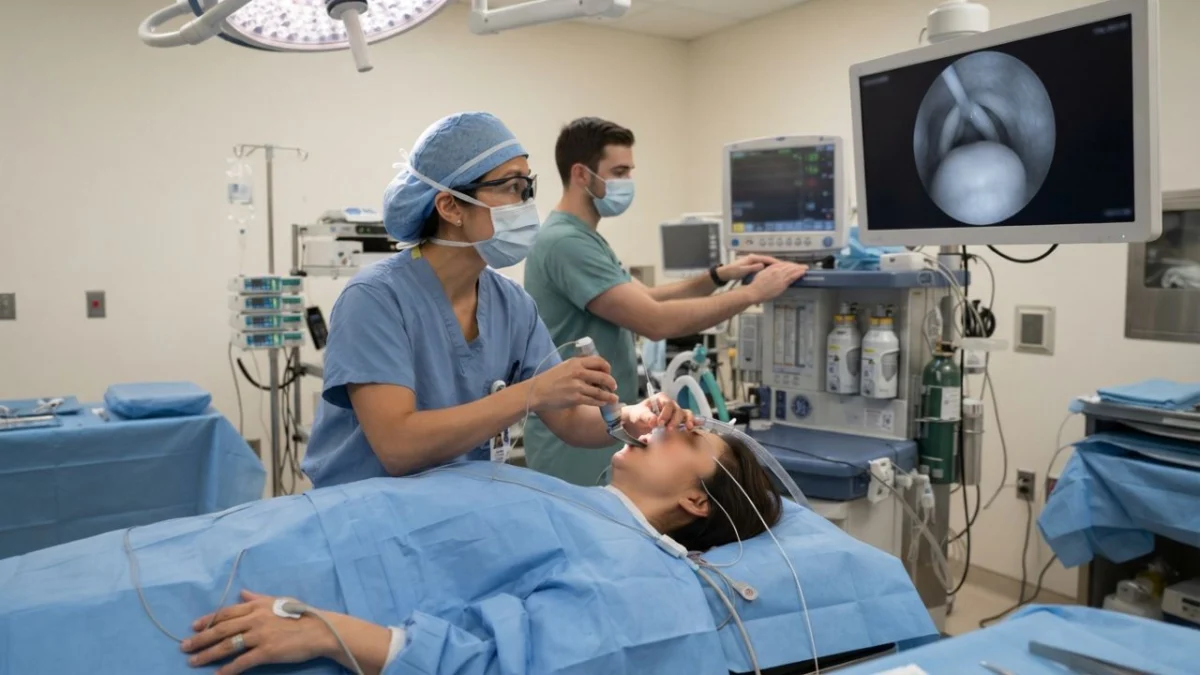
"A benign lump, invisible to standard preoperative checks, can transform a straightforward surgery into a life-threatening airway emergency when anesthesia relaxes the body."
Preoperative airway assessment, a cornerstone of safe anesthesia practice, typically relies on a combination of patient history, physical examination, and specific measurements. Clinicians meticulously inquire about neck mobility, the extent of mouth opening, jaw structure, and any history of snoring, hoarseness, or difficulty swallowing. These questions are designed to identify potential challenges in managing a patient’s airway, particularly during the critical moments of intubation. However, a recent case, detailed in a preprint by anesthesiologists at the Affiliated Yangming Hospital of Ningbo University, highlights a significant blind spot in this routine: asymptomatic epiglottic cysts.
The case involved a 60-year-old woman scheduled for laparoscopic gallbladder removal due to gallstones and cholecystitis. By all conventional preoperative assessments, her airway was deemed uncomplicated. She reported no subjective symptoms that would raise a red flag concerning her throat or breathing. Yet, upon induction of anesthesia, the anesthesiologist encountered immediate and severe difficulty with mask ventilation. Subsequent video laryngoscopy revealed the unexpected culprit: a large cyst on her epiglottis, a flap of cartilage at the base of the tongue that covers the opening of the larynx (the glottis). This cyst was so substantial that it completely obscured the glottis, the critical passage through which a breathing tube must be inserted to maintain ventilation. The first attempt at intubation, a procedure usually managed with practiced ease, failed.
This incident underscores a hidden category of anesthetic risk, one where a benign anatomical anomaly can pose a profound threat precisely because it remains undetected and asymptomatic until the patient is rendered unconscious. The cyst, a harmless presence in the awake state, becomes a formidable obstacle when the body’s natural muscle tone, which normally keeps the tongue and epiglottis away from the airway, is suppressed by anesthetic agents and muscle relaxants.
Epiglottic cysts, while uncommon, represent the most prevalent form of laryngeal cyst. They arise when the duct of a mucous gland within the epiglottis becomes blocked, leading to a buildup of glandular secretions. These cysts are benign and can manifest in individuals of any age. Historically, laryngoscopies reveal epiglottic cysts in approximately 4 out of every 5,000 procedures, equating to about 0.08%. Larger cysts, those more likely to cause obstruction, are even rarer, occurring in an estimated 0.02% of cases, according to research published in BMC Anesthesiology.
The danger of these cysts lies in their clinical presentation, or rather, their lack thereof. Congenital laryngeal cysts often present early in life with clear and dramatic symptoms, such as progressive airway obstruction, as seen in a case report of a five-day-old newborn whose cyst completely blocked his glottis. Acquired cysts in adults, however, frequently present a stark contrast. Many remain entirely asymptomatic, allowing them to grow undetected. As their size increases, some individuals may begin to experience subtle symptoms like a foreign body sensation in the throat, mild discomfort, or a feeling of obstruction. Significant breathing difficulties are typically only reported in advanced stages of growth, by which point the cyst has become considerably large. This insidious progression means that a patient may have a potentially airway-compromising lesion without having any reason to mention it during a preoperative assessment.
The critical moment when the danger emerges is during the induction of anesthesia. In a conscious individual, the inherent muscle tone in the tongue and pharynx plays a crucial role in maintaining a clear airway. This tone acts to keep the epiglottis and surrounding tissues positioned away from the laryngeal inlet. However, anesthetic drugs, particularly those used for induction and muscle relaxation, abolish this vital muscle tone. Consequently, the soft tissues of the upper airway can fall backward, and a cyst that was previously located on the epiglottis, without impinging on any structures, can then descend and obstruct the glottis.
Adding another layer of complexity, direct intervention to dislodge or remove the cyst during laryngoscopy carries its own significant risks. Attempting to push the cyst aside or manipulate it with a laryngoscope blade can inadvertently lead to its rupture. Clinicians writing in the Journal of Anaesthesiology Clinical Pharmacology have detailed the potentially catastrophic consequences of such an event. Rupture can result in the spillage of cyst fluid into the trachea, triggering a cascade of adverse reactions including laryngospasm (sudden, involuntary closure of the vocal cords), severe hypoxia (dangerously low blood oxygen levels), bradycardia (a slowed heart rate), and in the most severe scenarios, cardiac arrest.
The potential severity of these complications is illustrated by a peer-reviewed report in BMC Anesthesiology concerning a 61-year-old man. He was scheduled for elective arthroscopic surgery on his heel due to persistent pain. His preoperative airway assessment was exceptionally reassuring, with no prior history of difficult intubation and a Mallampati score of I, indicating an easily visualized airway. Following the administration of anesthetic agents, his airway pressure began to rise, and mask ventilation proved impossible. Attempts to improve ventilation by lifting the jaw were unsuccessful. Insertion of a laryngeal mask airway, a common rescue device, failed to establish adequate ventilation despite repeated adjustments. A second anesthesiologist was consulted, and a further intubation attempt using a light-guided technique also failed. It was only then that video laryngoscopy was employed, revealing a cyst measuring 2 by 3 centimeters that completely covered the epiglottis and obscured the vocal cords. An ear, nose, and throat surgeon was immediately summoned to prepare for an emergency tracheostomy. In a tense sequence that lasted approximately 15 minutes, the surgical team managed to use the tip of an endotracheal tube to lift the cyst, expose a portion of the glottis, and successfully pass a size 6.5 endotracheal tube through the narrow opening. During this critical period, the patient’s oxygen saturation briefly dropped to 85%.
In some instances, the cyst proves to be an insurmountable obstacle. A report in Cureus described an adult patient whose epiglottic cyst caused complete airway obstruction. Standard maneuvers, including positional changes of the larynx and blade techniques, were deemed too risky due to the high probability of cyst rupture. The team proceeded with a tracheostomy under local anesthesia. Even prior knowledge of a cyst does not guarantee a smooth anesthetic course. A 2023 report in Medicine detailed a patient specifically scheduled for the surgical removal of an epiglottic cyst. Despite this awareness, the patient experienced airway difficulties after induction of anesthesia when the cyst obstructed the glottis. In this case, a video laryngoscope ultimately facilitated successful intubation.
It is crucial to contextualize these reports. The Chinese preprint, being a single case and not yet peer-reviewed, cannot definitively establish the incidence of asymptomatic epiglottic cysts complicating anesthesia or the frequency of associated harm. Such statistics are difficult to ascertain precisely because these cysts are often discovered incidentally during procedures performed for unrelated reasons.
Neither the Chinese preprint nor the other case reports advocate for routine widespread screening of all patients’ airways with advanced imaging prior to elective surgery. Such an approach would be neither practical nor cost-effective. Instead, the consensus among clinicians and researchers points to more pragmatic conclusions. The fundamental importance of a comprehensive preoperative airway assessment cannot be overstated. Prompt utilization of advanced airway visualization tools like video laryngoscopy, rather than as a last resort after multiple failed attempts, is strongly recommended. Furthermore, the principle of early consultation with colleagues or specialists when faced with airway challenges remains paramount. The authors of the BMC Anesthesiology report emphasize that the advent of new technologies does not diminish the necessity of mastering traditional "front-of-neck" airway rescue techniques, such as cricothyroidotomy, which remain vital skills for managing unmanageable airways.
For patients, the most impactful action is straightforward yet specific. It involves proactive communication with the anesthesia team. Patients should be encouraged to report any throat-related symptoms, however seemingly trivial, at their preoperative anesthesia visit. This includes persistent sensations of something being stuck in the throat, any changes in voice quality, habitual snoring, or any difficulties experienced when swallowing. Additionally, individuals with a history of previous difficult intubation should explicitly disclose this information and inquire whether it has been adequately documented in their medical records. These details are invaluable for the anesthesia team in formulating a tailored anesthetic plan and mitigating potential risks. Ultimately, the decision-making process regarding anesthetic management rests with the experienced anesthesia team, who will weigh all available information to ensure patient safety.
