
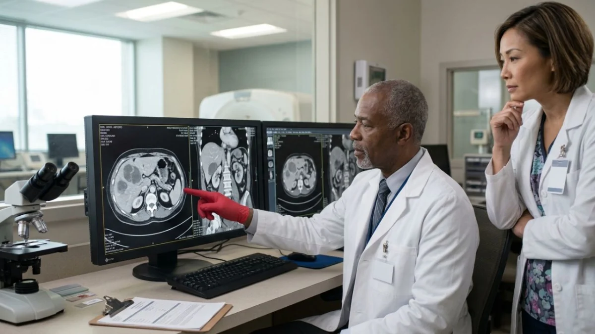
"When a patient’s scans scream ‘cancer,’ the subsequent biopsy can deliver a profound relief, or a stark reminder of medicine’s inherent complexities. This case underscores the critical role of histopathology in differentiating life-threatening malignancies from rare, benign conditions that can present with eerily similar radiological appearances."
In a remarkable medical case that highlights the intricate diagnostic challenges faced by clinicians, a 39-year-old woman presented with imaging findings so alarming they strongly suggested advanced cancer. Her liver was riddled with large, irregular masses, her abdominal lining dotted with small nodules, and her lymph nodes near the heart were enlarged. Radiologists, observing this pattern consistent with peritoneal carcinomatosis – the widespread spread of cancer throughout the abdominal lining – were prepared for a grim prognosis. However, a subsequent needle biopsy delivered a surprising and ultimately reassuring diagnosis: not cancer, but an inflammatory pseudotumor, a rare benign lesion capable of expertly mimicking malignancy on medical scans.
The perplexing case, detailed in the journal Oxford Medical Case Reports by radiologists from Avicenne Hospital at Mohammed V University in Rabat, Morocco, underscores a fundamental principle in medicine: imaging, while powerful, is often just the first step in a complex diagnostic journey. The woman, with no prior medical history, sought care for pain in her right lower back and upper middle abdomen. While she reported no fever, her physical examination revealed an enlarged and tender liver. Initial blood tests showed only minor liver enzyme elevations, with other key indicators like bilirubin, kidney function, and inflammatory markers remaining within normal limits. Crucially, tumor markers, which are often elevated in the presence of cancer, were not assessed at this stage.
The diagnostic odyssey began with an ultrasound, which revealed a misshapen liver suggestive of cirrhosis, several ill-defined lesions beneath its surface, and a small accumulation of fluid within the abdomen. The situation escalated with a contrast-enhanced computed tomography (CT) scan, a more detailed imaging modality. This scan unveiled multiple infiltrating masses. The most substantial mass, situated in the right lobe of the liver, measured an imposing 15 by 5 by 14 centimeters. This lesion was observed to encircle branches of the portal vein, the critical vessel that transports nutrient-rich blood from the intestines to the liver. While the portal vein remained patent, the mass abutted the gallbladder and duodenum without clear signs of invasion. Further examination of the CT scan revealed additional findings: nodules studding the liver’s surface, minute nodules scattered across the abdominal lining, a modest amount of ascites (abdominal fluid), and enlarged lymph nodes in the right cardiophrenic angle, a space nestled between the diaphragm and the heart. Some of these lesions exhibited central areas that appeared necrotic, or indicative of dead tissue. Taken in aggregate, these findings painted a compelling picture of widespread malignancy, a scenario that often necessitates aggressive treatment.
The turning point in this patient’s care came with a CT-guided needle biopsy, a procedure where a thin needle is used to extract a tissue sample from a suspicious lesion under imaging guidance. Under the microscope, pathologists examined the retrieved tissue. Instead of malignant cells, they observed a proliferation of fibroblasts and myofibroblasts – cells instrumental in wound healing and scar formation. These cells, lacking any abnormal features, were embedded within dense, scar-like tissue that was infiltrated by a variety of inflammatory cells. Critically, no cancer cells were identified. This detailed histological examination led to the definitive diagnosis of a hepatic inflammatory pseudotumor. It is important to note that the biopsy, by sampling only the lesion itself, could not definitively confirm or refute the presence of cirrhosis that had been suggested by the initial imaging.
The authors of the report acknowledged certain limitations in the diagnostic process. Crucially, tests to detect ALK (anaplastic lymphoma kinase), a protein marker that can help differentiate inflammatory pseudotumors from a related but distinct entity known as an inflammatory myofibroblastic tumor, were not performed. Similarly, testing for IgG4, an immunoglobulin that can be associated with certain immune-mediated conditions that might contribute to inflammatory processes, was also absent. Furthermore, the patient was lost to follow-up after her diagnosis, meaning her long-term outcome – whether the lesions naturally regressed, grew, or required intervention – remains unknown. This lack of follow-up is a common challenge in medical research and clinical practice, highlighting the importance of robust patient engagement and tracking.
Inflammatory pseudotumors are a fascinating group of lesions that can arise in various organs, but they are most commonly encountered in the lungs. Their occurrence within the liver is considered rare. The clinical presentation of these pseudotumors can be varied; approximately 15.6% of affected individuals are asymptomatic. A review from 2011 underscored the propensity of hepatic inflammatory pseudotumors to be misdiagnosed as cancer, a testament to their deceptive radiological appearance. The precise etiology of these lesions remains elusive, with proposed triggers encompassing infections, autoimmune dysregulation, and chronic liver or bile duct diseases.
A multicenter study involving 45 patients treated across three South Korean hospitals between 1995 and 2011 provided further insight into these challenging diagnoses. This research indicated that while both CT and magnetic resonance imaging (MRI) could offer suggestive clues, they lacked definitive features to distinguish pseudotumors from malignancy. Consequently, tissue diagnosis through biopsy was deemed essential. In this cohort, tumor markers were notably normal in an overwhelming 93.3% of patients. Ten patients underwent surgical resection, and importantly, none experienced a recurrence, suggesting the benign nature of these lesions once correctly identified.
The presence of peritoneal involvement, as seen in this particular case, amplifies the diagnostic dilemma. The authors noted only a limited number of published cases describing hepatic pseudotumors with concomitant lesions in the peritoneum or omentum, the fatty apron that drapes over the abdominal organs. This rarity further contributes to the diagnostic confusion. The presence of underlying liver disease, such as cirrhosis, adds another layer of complexity. Individuals with cirrhosis face an elevated risk of developing primary liver cancer, as recognized by the National Cancer Institute. Therefore, the discovery of an unusual mass within a liver exhibiting cirrhotic changes is, understandably, treated with a high degree of suspicion for malignancy.
The central role of histopathology, the microscopic examination of tissue, cannot be overstated. The authors emphasized that "Image-guided biopsy is recommended for indeterminate lesions to establish diagnosis and avoid unnecessary surgery." This principle is paramount, particularly when the stakes are as high as distinguishing between a treatable benign condition and a potentially life-threatening cancer.
Once cancer is definitively ruled out, the management of hepatic inflammatory pseudotumors is not standardized. The natural history of these lesions can vary; some may spontaneously regress, while others may respond to non-surgical interventions. Reports suggest that anti-inflammatory medications or antibiotics can be effective in certain cases. In the aforementioned Korean study, a significant proportion of patients (35 out of 45) were managed conservatively without surgery. Of these, 20 received antibiotic therapy, and remarkably, all patients who underwent follow-up imaging demonstrated either shrinkage or complete resolution of their lesions.
While this case offers a profoundly positive outcome for the patient, it is crucial to avoid drawing sweeping conclusions. The presence of suspicious liver scans does not invariably portend a benign diagnosis. Imaging findings that strongly resemble spreading cancer necessitate urgent and thorough evaluation. However, this case powerfully illustrates why obtaining a tissue diagnosis is an indispensable step before embarking on major treatment decisions. For patients confronting concerning imaging results, engaging with their healthcare team to understand the potential role of a biopsy or a specialist consultation is a prudent measure before commencing any therapeutic regimen. This detailed understanding empowers patients and ensures that the most appropriate and effective course of action is pursued, based on definitive diagnostic evidence rather than solely on the suggestive, albeit powerful, evidence from medical imaging. The case serves as a compelling reminder that while technology advances, the foundational principles of medical diagnosis, grounded in meticulous observation and tissue analysis, remain indispensable.
