
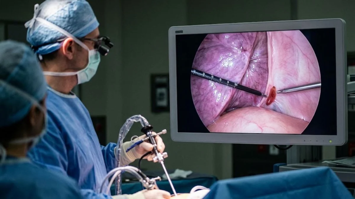
"Observing a living adult Paragonimus worm moving during a minimally invasive chest procedure marks a significant advancement in diagnosing a notoriously elusive parasitic infection, often mistaken for tuberculosis or lung cancer."
The diagnosis of paragonimiasis, an infection caused by lung flukes, has historically been a diagnostic challenge, often leading to prolonged periods of misdiagnosis and ineffective treatment. A recent case, detailed in Infection, Genetics and Evolution, has provided unprecedented visual confirmation of a live adult Paragonimus worm within the pleural cavity of a patient. This direct visualization, achieved through thoracoscopy, offers a tangible leap forward in understanding and potentially diagnosing this rare but serious parasitic disease, which frequently mimics other more common pulmonary conditions. The findings underscore the importance of considering rare parasitic infections in differential diagnoses, especially in patients with specific exposure histories and perplexing symptoms.
A Glimpse of the Elusive Parasite: A Case of Direct Visualization
Doctors rarely encounter the lung fluke responsible for paragonimiasis. Typically, diagnosis relies on identifying microscopic eggs in sputum or pleural fluid, a method that is often unreliable and can fail to detect the parasite, leading to delayed diagnosis and months of incorrect treatment for affected patients. However, in a remarkable case involving a 38-year-old woman experiencing recurrent pleural effusion, the diagnostic question was settled instantly when a thoracoscope revealed a living adult Paragonimus worm on the right posterior pleura near the costal angle. The worm’s peristaltic, wave-like motion provided irrefutable evidence of its presence. This unprecedented direct visualization of a living adult Paragonimus under thoracoscopy, as reported by the study’s authors, represents a significant milestone in the diagnostic capabilities for this parasitic infection.
The woman’s medical journey was marked by two hospitalizations within a six-month period due to pleural effusion, the accumulation of fluid in the space between the lung and the chest wall. During her initial admission, serological testing, specifically an ELISA, indicated the presence of antibodies against Schistosoma japonicum and sparganum, two other parasitic species. Based on these results, a parasitic infection was suspected, and praziquantel, a broad-spectrum antiparasitic medication, was administered.
Despite the initial treatment, the pleural effusion recurred, necessitating a second admission. This time, physicians opted for a medical thoracoscopy, a minimally invasive surgical procedure that allows direct visualization of the chest cavity. It was during this procedure that the offending worm was discovered. Subsequent microscopic examination identified the parasite as Paragonimus. Further confirmation was obtained through metagenomic next-generation sequencing of the pleural fluid, which also detected Paragonimus, providing two independent lines of evidence for the diagnosis. Following this definitive diagnosis, the patient received another course of praziquantel. At a four-month follow-up, the pleural effusion had not returned, indicating successful treatment.
This sequence of events highlights a critical challenge in parasitic disease diagnosis: antibody cross-reactivity. Positive serological results for one fluke species do not definitively confirm the specific type of fluke responsible for the infection. In this case, the initial treatment, while seemingly reasonable given the serological findings, was ultimately aimed at the wrong parasite. This underscores the diagnostic dilemma physicians face when faced with ambiguous serological data and the crucial role of definitive diagnostic methods when available.
Understanding Paragonimus: The Lifecycle and Transmission
Paragonimus is a genus of trematodes, commonly known as lung flukes. Humans typically acquire infection by consuming raw or undercooked freshwater crabs and crayfish that harbor the larval stages of the parasite. Several species of Paragonimus can infect humans, with Paragonimus westermani being one of the most widely distributed globally.
The lifecycle of the Paragonimus parasite is intricate. After ingestion, the larvae excyst in the small intestine and penetrate the duodenal wall. They then migrate through the peritoneal cavity for approximately a week before traversing the diaphragm to enter the pleural cavity. From there, they move through lung tissue, eventually reaching the bronchi, where they mature into adult worms within cystic cavities. This maturation process takes about five to six weeks. Adult flukes are reddish-brown and typically measure between 7.5 to 12 millimeters in length.
A significant diagnostic hurdle arises from the fact that adult flukes do not produce eggs until they reach maturity. This creates a critical window of more than a month after infection during which sputum testing, a common diagnostic method, will yield no positive results. Even after egg production begins, detection can be unreliable, particularly in cases of light infections. This is precisely why antibody testing is employed, and it also explains how this patient’s initial serology directed her physicians toward an incorrect diagnosis.
While often associated with Asia, paragonimiasis is not geographically confined. A published case series from the United States described four American patients, three of whom had consumed live crabs at sushi bars, with one instance involving a crab served in a martini. In North America, Paragonimus kellicotti is the species most commonly implicated. The Centers for Disease Control and Prevention (CDC) guidance notes that several cases in the U.S. have been linked to river raft float trips in Missouri where uncooked crawfish were consumed.
Mimicking the Dreaded: Paragonimiasis vs. Tuberculosis and Cancer
The clinical presentation of paragonimiasis often includes a persistent cough, chest pain, blood-tinged sputum, and, frequently, eosinophilia – an elevated count of a specific type of white blood cell that plays a role in allergic reactions and parasitic infections. Chest imaging can reveal a range of abnormalities, including cavities, nodules, patchy consolidation, and effusions.
This constellation of symptoms and imaging findings closely resembles tuberculosis (TB), a highly prevalent infectious disease. Consequently, patients in TB-endemic regions have often been treated with anti-tuberculous drugs before the possibility of parasitic infection is reconsidered. In one documented Korean case, a 38-year-old man underwent two months of empirical TB treatment before surgery, during which Paragonimus eggs were finally discovered in his pleura.
The complications of paragonimiasis can be severe. A study involving 179 patients with paragonimiasis at a Korean university hospital reported that 10.6% developed pneumothorax, or a collapsed lung. Notably, asthma was found to be significantly more common in patients who subsequently developed pneumothorax, suggesting a potential underlying predisposition or interaction.
Fortunately, the treatment for paragonimiasis is relatively straightforward and effective once the diagnosis is established. Praziquantel is the drug of choice, administered at a dosage of 25 milligrams per kilogram of body weight, three times daily for two consecutive days. Patients typically experience rapid improvement following treatment. The primary challenge, therefore, lies not in the treatment itself, but in achieving an accurate diagnosis in the first place.
Implications for Consumption of Freshwater Crustaceans
While this single case report is a significant medical event, it does not suggest that lung fluke infection is a common consequence of consuming seafood. Paragonimiasis remains a neglected tropical disease, and most clinicians in the United States will likely never encounter a patient with this condition during their careers.
However, the route of exposure is specific and, crucially, avoidable. The risk is primarily associated with consuming raw or undercooked freshwater crabs and crayfish. It is important to note that traditional preparation methods such as marinating crab in vinegar, brine, or wine without adequate cooking do not reliably kill the infective larvae. Similarly, salting, pickling, or lightly smoking these crustaceans is insufficient to eliminate the risk. Thorough cooking, on the other hand, is effective. The CDC recommends heating crabs and crayfish to an internal temperature of at least 145 degrees Fahrenheit to ensure the destruction of Paragonimus larvae.
From a practical standpoint, the key takeaway for patients is the importance of a detailed medical history that includes potential exposures. An individual presenting with a persistent cough, chest pain, blood in their sputum, and an elevated eosinophil count, particularly if they have a history of consuming raw freshwater crustaceans or have spent time in an endemic region, should explicitly disclose this information to their healthcare provider. This seemingly minor detail can fundamentally reframe the diagnostic workup.
It is also imperative that individuals experiencing such symptoms consult a clinician for evaluation rather than self-diagnosing a parasitic cause. The clinical presentation of paragonimiasis can overlap with numerous other conditions, many of which are far more common. A comprehensive medical assessment is essential to differentiate between these possibilities and arrive at the correct diagnosis.
Key Questions Answered About Paragonimiasis
What is paragonimiasis?
Paragonimiasis is an infection caused by parasitic lung flukes belonging to the genus Paragonimus. It is acquired through the consumption of raw or undercooked freshwater crabs and crayfish and typically resides in the lungs.
What made this case unusual?
This case is unusual because doctors directly observed a living adult Paragonimus worm moving on the pleural lining during a thoracoscopy. Diagnosis in most cases relies on finding microscopic eggs, and this direct visualization is described as a first in medical literature.
How is it usually diagnosed?
Diagnosis is typically made by identifying microscopic Paragonimus eggs in sputum samples. Less commonly, eggs may be found in stool or pleural fluid. Antibody testing can aid in diagnosis, but the flukes do not produce eggs until they mature, which can take approximately five to six weeks after infection.
Why is it so often missed?
Paragonimiasis is frequently missed because its symptoms and chest imaging findings closely resemble those of tuberculosis and lung cancer. As a result, patients have sometimes been treated for TB for extended periods before the parasitic cause is identified.
Can this happen in the United States?
While rare, paragonimiasis cases have been reported in the United States. These typically involve Paragonimus kellicotti and are often linked to the consumption of raw crayfish or live crabs obtained from restaurants.
How is it treated?
The drug of choice for treating paragonimiasis is praziquantel. It is administered at a dosage of 25 milligrams per kilogram of body weight, three times daily for two consecutive days. Most patients experience a rapid improvement in their symptoms once the correct diagnosis is made and treatment is initiated.
