
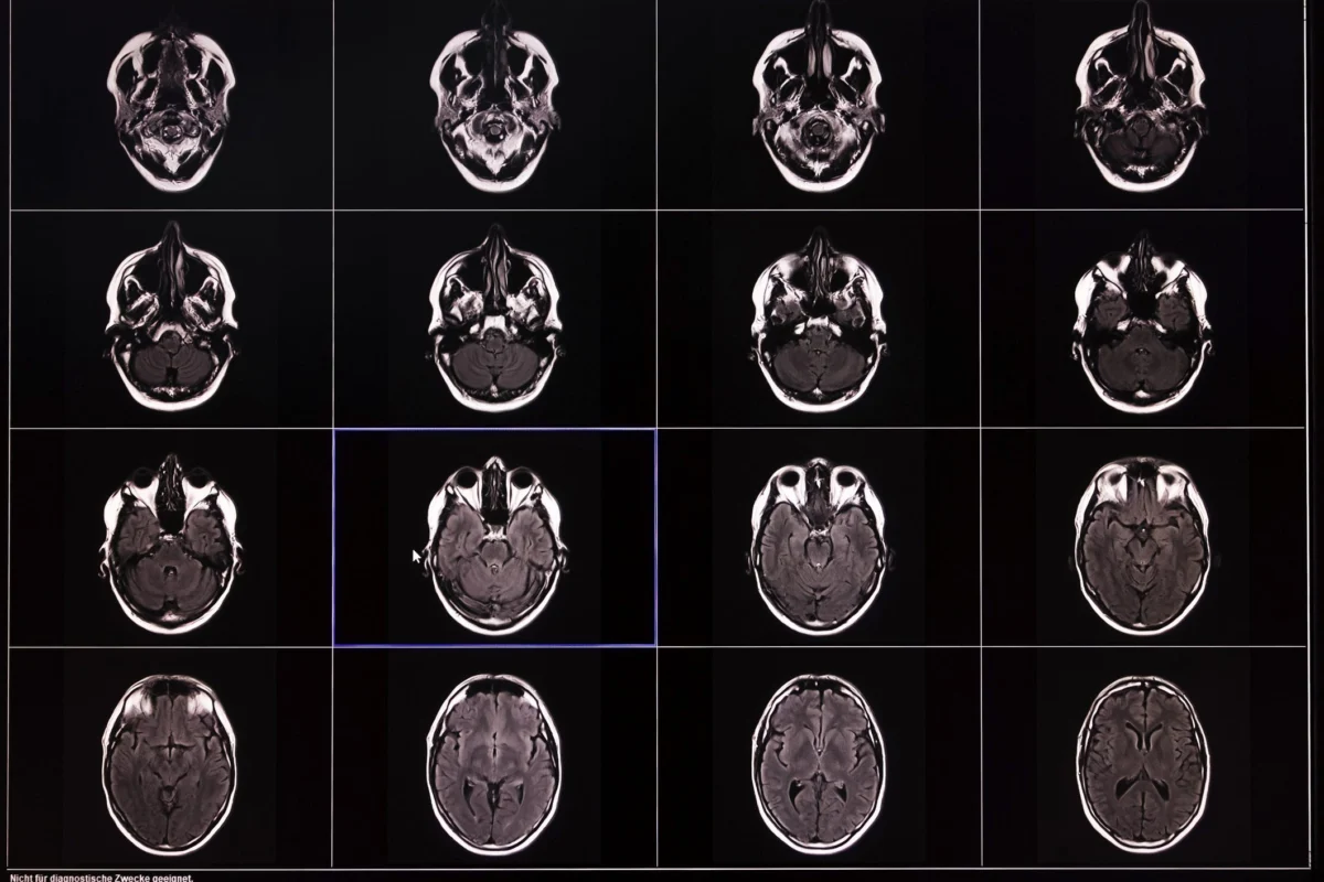
"Memory decline in aging is not the result of a single regional failure, but rather a reflection of a broad biological vulnerability in brain structure that accumulates across multiple interconnected networks over several decades."
While the hippocampus has long been the primary focal point in the study of age-related memory loss and Alzheimer’s disease, a landmark analysis of over 10,000 brain scans suggests this narrow focus may be insufficient. By integrating data from dozens of research cohorts, neurologists have discovered that the structural changes responsible for cognitive decline are far more distributed than previously understood. This shift in perspective moves the scientific community away from a "single-site" theory of memory toward a "whole-brain" model, where the degradation of visual, auditory, and connective tissues plays a role as critical as the hippocampus itself in determining how well we remember as we age.
The Power of Big Data in Neurology
The study, recently published in the journal Nature Communications, represents one of the most comprehensive efforts to map the aging brain to date. To reach their conclusions, a team of researchers, including senior author Dr. Alvaro Pascual-Leone, a professor of neurology at Harvard Medical School, analyzed a massive dataset consisting of 10,343 MRI scans. This structural data was cross-referenced with 13,460 memory test results from 3,737 cognitively healthy adults.
In the past, neurological studies were often limited by small sample sizes, which can lead to "noise" or over-emphasize the importance of specific brain regions. By using a "big data" approach, researchers were able to filter out individual anomalies and identify universal patterns of decay. This allowed them to construct a high-resolution timeline of how the human brain changes from mid-life into old age, providing a clearer picture of why some individuals maintain sharp cognitive function while others experience rapid decline.
Moving Beyond the Hippocampus
For decades, the hippocampus—a small, seahorse-shaped structure located deep within the temporal lobe—has been the "protagonist" of memory research. It is the primary engine for learning and the storage of new information. Because it is often the first area to show significant damage in patients with Alzheimer’s disease, it became the gold standard for measuring brain health.
However, the findings in Nature Communications suggest that focusing solely on the hippocampus is like looking at a single engine part while ignoring the rest of the vehicle. While the study confirmed that the hippocampus does indeed undergo significant structural changes—often beginning as early as a person’s late 50s—it also revealed that this is only one piece of a much larger puzzle. The researchers identified four additional brain areas with direct connectivity to the hippocampus that also showed significant volume loss.
This discovery highlights the importance of "neural highways." As we age, the connections that allow the hippocampus to communicate with the rest of the brain begin to weaken. When these pathways fray, the brain loses its ability to integrate information effectively, even if the hippocampus itself remains relatively intact.
The Role of the Temporal Lobe and Sensory Processing
One of the most significant revelations of the study was the involvement of six other brain regions located within the temporal lobe. This area of the brain is primarily responsible for processing sensory input—specifically auditory and visual information.
The authors suggest that the atrophy of these regions creates a "bottleneck" for memory. Memory is not an abstract concept; it is built from the sights and sounds of our daily lives. If the brain’s ability to process visual and auditory information is compromised, the "raw materials" needed to create a memory are degraded before they even reach the hippocampus. For example, if the regions responsible for visual detail begin to shrink, a person may find it harder to recall the specific features of a face or the layout of a room, not because their "storage" is full, but because the "input" was never clearly processed.
This suggests that memory loss may often begin as a sensory processing issue. When the temporal lobe regions responsible for environmental interaction lose volume, the details of a memory become fuzzy, leading to the "tip-of-the-tongue" moments and general forgetfulness often associated with aging.

The Cumulative Effect: Why Some Age Faster Than Others
The study provides a new framework for understanding the pace of cognitive decline. Rather than viewing memory loss as a steady, inevitable slide, the researchers found that decline is often accelerated by the number of affected regions.
Everyone experiences some degree of brain aging, but the threshold for "cognitive disability" is reached when multiple areas begin to shrink simultaneously. The more regions that show volume loss—and the more those regions are interconnected—the faster a person’s memory tends to fail. This explains why two people of the same age can have vastly different cognitive abilities: one may have localized shrinkage that the brain can compensate for, while the other may have a systemic "thinning" across several critical networks.
Dr. Pascual-Leone notes that this reflects a "broad biological vulnerability." This vulnerability is likely influenced by a combination of genetics, lifestyle, and environmental factors that accumulate over a lifetime. It isn’t just about one "memory gene"; it is about how the entire organ resists the cumulative wear and tear of decades of life.
Clinical Implications and Early Intervention
The shift toward a multi-regional understanding of the brain has profound implications for the future of medicine. Currently, many diagnostic tools and potential treatments for dementia focus heavily on the hippocampus. By expanding the "target zone" to include the temporal lobe and connective networks, researchers can develop more sensitive screening processes.
"Understanding this can help researchers identify individuals at risk early," Dr. Pascual-Leone explains. If doctors can detect volume loss in the temporal lobe or connectivity networks before the hippocampus shows signs of decay, they may be able to intervene years earlier. This opens the door for "precision neurology"—personalized interventions designed to support the specific areas of an individual’s brain that are most vulnerable.
Strategies for Cognitive Longevity
While the study paints a complex picture of brain aging, it also reinforces the idea that the brain is a dynamic, plastic organ. The concept of "neuroplasticity" suggests that the brain can reorganize itself and form new connections to compensate for tissue loss in other areas. To foster this resilience, experts recommend a multi-pronged approach to brain health.
1. Physical Movement and Blood Flow:
Physical exercise is perhaps the most effective way to maintain brain volume. Aerobic exercise increases blood flow to the brain and stimulates the release of brain-derived neurotrophic factor (BDNF), a protein that supports the survival of existing neurons and encourages the growth of new ones. Regular movement has been shown to specifically preserve volume in the hippocampus and the surrounding temporal regions.
2. Cognitive Engagement and Novelty:
The "use it or lose it" principle applies directly to neural networks. Engaging the brain through learning new skills—such as a foreign language, a musical instrument, or a complex hobby—forces the brain to build new pathways. This "cognitive reserve" acts as a buffer; even if some areas of the brain begin to shrink, the person can maintain high function because they have a wealth of alternative neural routes to draw upon.
3. Sensory Maintenance:
Given the study’s findings on the temporal lobe, maintaining sensory health is crucial. Treating hearing loss and vision problems is not just about quality of life; it is about ensuring the brain continues to receive the high-quality input it needs to form and store memories. Research has consistently shown a link between untreated hearing loss and an increased risk of dementia, likely due to the resulting atrophy in auditory processing regions.
A New Era of Brain Health
The transition from a hippocampus-centric view to a whole-brain perspective marks a significant milestone in neurology. It acknowledges the complexity of the human mind and the intricate dance between different brain regions. As we move forward, the focus will likely shift from "curing" a single region to "fortifying" the entire system.
By understanding that memory decline is a decades-long process involving multiple structural changes, we gain the power to act earlier. Through a combination of medical screening and proactive lifestyle choices, it is becoming increasingly possible to not only understand the aging brain but to actively protect the networks that make us who we are.
