
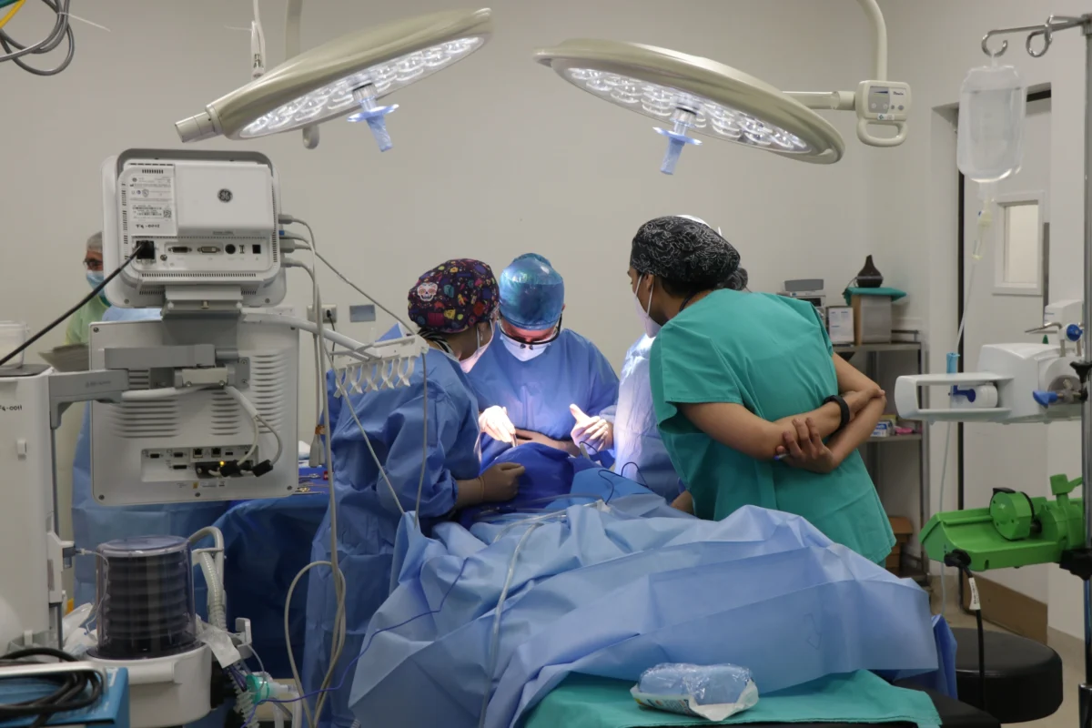
"In a groundbreaking development, a baby born with gastroschisis has undergone a successful pioneering operation while still in the womb, marking a significant milestone in fetal surgery and offering a beacon of hope for improving outcomes in complex congenital conditions."
This remarkable case involves a newborn, Theo, who was diagnosed with gastroschisis, a rare and serious birth defect. In a departure from traditional treatment, which typically involves surgery after birth, Theo’s medical team opted for an innovative in-utero procedure designed to protect his exposed intestines. This pioneering approach, performed during pregnancy, has yielded positive results, with both mother and baby recovering well. The success of this intervention not only represents a significant advancement in prenatal medicine but also opens new avenues for managing gastroschisis, potentially transforming the prognosis for affected infants and their families.
Gastroschisis is a congenital anomaly characterized by a defect in the abdominal wall, usually to the right of the umbilical cord, through which the intestines, and sometimes other abdominal organs, protrude. Unlike omphalocele, another condition involving externalized organs, gastroschisis lacks a protective membrane, leaving the delicate bowel tissue vulnerable to damage from exposure to amniotic fluid within the uterus. This exposure can lead to inflammation, shortening, and other complications that can significantly impact the infant’s health and require extensive treatment after birth.
The exact etiology of gastroschisis remains elusive, though current research suggests a complex interplay of genetic predispositions and environmental influences. Factors such as younger maternal age have been identified as a risk factor, but the condition can manifest in any pregnancy. Diagnosis is typically made during routine prenatal screening through ultrasound, which can identify the characteristic abdominal wall defect. Elevated maternal levels of alpha-fetoprotein (AFP), a protein produced by the fetus, can also serve as an indicator, prompting further investigation.
Historically, infants born with gastroschisis have faced a challenging road, often requiring immediate surgical intervention shortly after birth. The primary goal of this surgery is to carefully reposition the extruded organs back into the abdominal cavity and to close the abdominal wall defect. The complexity of the surgery and the subsequent recovery period can vary significantly depending on the extent of the defect and the condition of the exposed bowel. In some cases, a single operation may suffice, while in others, a staged approach over several days or even weeks might be necessary to gradually return the organs to their rightful place and allow the abdominal cavity to accommodate them. This can involve specialized techniques such as silo placement, where a protective bag is used to temporarily house the organs before they are reduced.
The groundbreaking procedure performed on Theo represents a paradigm shift in the management of gastroschisis. Instead of waiting for birth to address the condition, surgeons intervened while the fetus was still developing within the protective environment of the womb. This fetal surgery was meticulously planned and executed after prenatal scans clearly identified the gastroschisis. The primary objective was to shield the exposed intestines from the potentially damaging effects of prolonged contact with amniotic fluid. By performing this intervention in utero, the medical team aimed to mitigate the inflammation and injury that can occur during the latter stages of pregnancy, thereby potentially improving the overall health of the bowel at the time of birth and simplifying subsequent postnatal care.
Following the in-utero procedure, Theo’s mother, Maisie Savage, and the baby were subjected to close medical monitoring throughout the remainder of the pregnancy. This meticulous surveillance was crucial to ensure the well-being of both mother and fetus and to prepare for the delivery. The successful recovery of both Maisie and Theo post-delivery has been widely reported, generating considerable international attention within the medical community. This case is being hailed as an important proof of concept for fetal surgery in carefully selected patients diagnosed with gastroschisis. However, specialists are quick to emphasize that while this represents a significant leap forward, additional research and clinical trials are indispensable to fully understand the safety, efficacy, and long-term benefits of this approach. Determining which specific patient profiles are most likely to benefit from such prenatal intervention will be a key focus of future studies.
The implications of this pioneering surgery are far-reaching. It underscores the remarkable advancements being made in the field of maternal-fetal medicine and pediatric surgery. The ability to diagnose and, in select cases, treat congenital conditions before birth opens up unprecedented possibilities for improving infant health outcomes. Prenatal imaging technologies have become increasingly sophisticated, allowing for earlier and more accurate diagnoses. Coupled with the development of refined surgical techniques, physicians are now empowered to intervene in ways that were once considered science fiction. This proactive approach aims to minimize the severity of conditions, reduce the need for extensive postnatal interventions, and ultimately enhance the quality of life for children born with complex medical challenges.
While the experimental nature of this fetal surgery means it is unlikely to replace standard postnatal surgical management in the immediate future, its apparent success offers a compelling glimpse into the future of treating gastroschisis. The hope is that with further validation, this in-utero approach could become a more integrated part of the therapeutic arsenal for certain pregnancies affected by this condition. This would not only offer a more favorable prognosis for affected infants but also provide a greater sense of empowerment and hope for families facing such a diagnosis.
The journey from diagnosis to recovery for infants with gastroschisis has traditionally been arduous. However, cases like Theo’s illuminate the transformative potential of continuous medical research and innovation. The rigorous evaluation of novel procedures is paramount to ensure they meet the highest standards of safety and efficacy for both mother and child. If subsequent clinical investigations confirm the promise demonstrated in this unique case, fetal surgery for gastroschisis could significantly expand the spectrum of treatment options available, offering a more optimistic outlook for many families.
Beyond the immediate benefits for infants with gastroschisis, this successful outcome serves as a powerful testament to the broader potential of advancements in maternal-fetal medicine. By addressing critical health issues before birth, these developments have the capacity to profoundly impact infant survival rates, reduce the incidence of long-term complications, and foster improved developmental trajectories for children born with a wide array of complex congenital conditions. The ongoing pursuit of knowledge and the translation of scientific discoveries into clinical practice are vital to continuing this progress and ensuring that future generations of babies have the best possible start in life. The case of Theo Savage stands as a beacon, illuminating the path forward in the ever-evolving landscape of prenatal care and fetal intervention.
