
"A simple alcohol swab can unlock a diagnosis, potentially saving patients from unnecessary invasive procedures for a harmless skin condition that often masquerades as stubborn dirt."
A recent case report published in the Indian Journal of Dermatology, Venereology and Leprology has illuminated a less common presentation of terra firma-forme dermatosis (TFFD), a benign skin condition characterized by a buildup that resembles dirt but resists conventional washing. This particular instance involved a 27-year-old woman whose skin lesions were not the typical flat, dirt-like patches but rather raised, rough, and follicle-centered bumps. This atypical presentation highlights the diagnostic challenges posed by TFFD and underscores the importance of a simple, yet effective, diagnostic test that can swiftly differentiate it from more serious dermatological concerns. The case, meticulously documented by a team of dermatologists from N.K.P. Salve Institute of Medical Sciences and Research Centre and Lata Mangeshkar Hospital in Nagpur, introduces what the authors have termed a new "folliculo-papillomatous variant" of TFFD, expanding the known clinical spectrum of this intriguing condition.
Terra firma-forme dermatosis, first described in 1987 by Duncan and colleagues and sometimes colloquially referred to as "Duncan’s dirty dermatosis," is a benign cutaneous condition that presents as a deposit on the skin. Its hallmark is its resistance to ordinary soap and water cleansing, leading many to believe it is simply ingrained dirt. Typically, TFFD manifests as flat, brown patches that can appear on various parts of the body, including the neck, face, trunk, ankles, and navel. However, the case in question deviated significantly from this typical presentation. The patient’s lesions were not flat but raised, exhibiting a rough texture. Crucially, they were clustered and centered around hair follicles, with some coalescing to form intricate, lacy, netlike plaques. These eruptions had been present for approximately two months, were non-itchy, and caused no discomfort.
The initial assessment by the dermatologists involved considering a differential diagnosis that encompassed several possibilities. Among the conditions under consideration were fungal infections, which often present with scaling and discoloration, and the rare papillomatosis disorders, which can manifest with similar netlike or papillomatous patterns. The patient’s presentation was complex enough to warrant a thorough investigation. Preliminary tests, such as a potassium hydroxide (KOH) examination to detect fungal elements, returned negative results, ruling out common fungal culprits like pityriasis versicolor, a prevalent yeast infection that can cause discolored patches. The team also considered confluent and reticulated papillomatosis, a rare genodermatosis characterized by a net-like pattern of papules, often on the trunk.
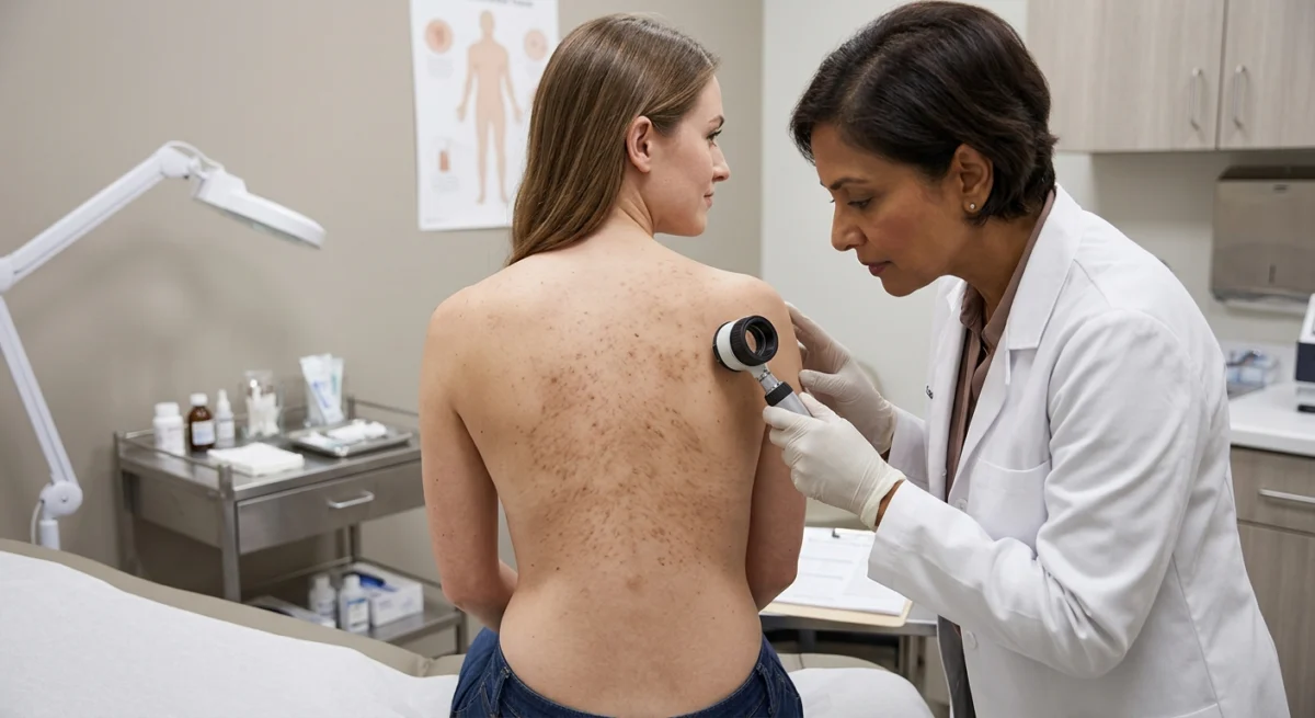
A pivotal moment in the diagnostic process arrived when the dermatologists employed a simple yet highly effective maneuver: wiping the affected skin with an alcohol swab. This action led to the removal of the patches, a crucial clue that pointed towards TFFD. Under dermoscopy, a non-invasive imaging technique that magnifies skin structures, the physicians observed characteristic findings. These included brown clumps aggregated around hair follicles, areas where the follicles themselves appeared spared, and an exaggerated reticulated pattern of pigmentation. The combination of these dermoscopic observations and the prompt clearing of the lesions with alcohol unequivocally confirmed the diagnosis of this novel variant of terra firma-forme dermatosis.
The diagnostic utility of the alcohol swab test is a cornerstone in identifying TFFD, particularly in its atypical presentations. The Indian Journal of Dermatology, Venereology and Leprology case report emphasizes that this simple procedure can often obviate the need for more extensive and invasive diagnostic workups. A stepwise approach, as suggested by authors in CosmoDerma, is recommended. Initially, washing with soap and water should be attempted. This step is vital for differentiating TFFD from dermatosis neglecta, a condition that arises from inadequate cleansing and typically resolves with thorough hygiene. If soap and water prove ineffective, the next step is the application of 70% isopropyl alcohol. The swift and near-complete resolution of the lesions upon alcohol application is considered diagnostic for TFFD. Failure of both these methods should prompt further investigation into alternative diagnoses.
The implications of this simple diagnostic approach are significant. It can prevent patients from undergoing unnecessary procedures, such as punch biopsies. A case reported in Cureus in January 2026 illustrated this point. A 25-year-old man presented with a persistent, wart-like yellowish plaque on his chest. He underwent a punch biopsy, which revealed thickened, compacted outer skin layers without significant inflammation—findings consistent with TFFD. However, prior to the biopsy, an alcohol wipe had produced immediate and near-complete clearing of the lesion. This highlights how a simple alcohol test could have spared the patient the discomfort and cost of a biopsy. TFFD is frequently misdiagnosed and confused with other conditions such as acanthosis nigricans, characterized by velvety darkening of skin folds, and "dirty neck" pigmentation, which is a form of dermatosis neglecta.
Despite its appearance, TFFD is not an indicator of poor personal hygiene. The underlying pathophysiology is believed to involve a disorder of keratin retention, stemming from abnormal maturation and shedding of skin cells. According to DermNet, a comprehensive online resource for dermatological information, a delay in the normal shedding process leads to the trapping of melanin (skin pigment), sebum (natural skin oil), and microorganisms within the stratum corneum, the outermost layer of the epidermis. This entrapment creates the characteristic dirty appearance.
Several factors have been implicated as potential contributors to this abnormal keratin retention. The CosmoDerma authors suggest that heavy use of occlusive creams, prolonged sun exposure, and colonization by Malassezia yeast, a common inhabitant of the skin, may play a role. Furthermore, a link with atopic dermatitis (eczema) has been proposed. In individuals with atopic dermatitis, a compromised skin barrier and excessive use of emollients might impair the natural desquamation process, leading to the accumulation of skin debris. The CosmoDerma case study involved a 5-year-old girl with atopic dermatitis who used moisturizers extensively. Her mother reported that conventional treatments, including soap, water, and topical steroids, had failed to resolve the skin discoloration.
The authors of the Indian Journal of Dermatology, Venereology and Leprology report argue that TFFD is likely underreported and more prevalent in clinical practice than current literature suggests. They emphasize that while the condition is benign, its tendency to be misdiagnosed, especially in its atypical forms, is a significant concern. The recognition of variants such as the warty, papillomatous, and netlike presentations is crucial for dermatologists to ensure accurate and timely diagnosis. Similarly, the CosmoDerma authors underscore the issue of misdiagnosis due to the condition’s rarity and its resemblance to other pigmentary disorders, leading to inappropriate treatments.
The identification of unusual presentations, like the folliculo-papillomatous variant described in the recent case, can prompt clinicians to consider the simple alcohol test earlier in the diagnostic pathway. This can expedite diagnosis and prevent the initiation of ineffective or potentially harmful treatments. Advances in dermatological tools are also aiding in the identification of TFFD. For instance, ultraviolet-induced fluorescence dermoscopy is being explored as an additional method to help diagnose the condition. In classic cases, dermoscopy might reveal characteristic platelike brown scales arranged in a cobblestone pattern, although this finding is not universally present.
It is important to note that the current understanding of TFFD is largely based on individual case reports and series, which, while valuable, do not allow for definitive conclusions about the condition’s incidence or prevalence. Furthermore, dark or changing skin patches can arise from a multitude of causes, some of which necessitate prompt medical intervention. Therefore, self-diagnosis based on the appearance of skin lesions is strongly discouraged. Individuals experiencing new, spreading, or changing pigmentation should seek professional evaluation from a dermatologist or primary care physician to ensure an accurate diagnosis and appropriate management. The advent of new variants and the persistent challenge of misdiagnosis highlight the ongoing need for increased awareness and refined diagnostic strategies for terra firma-forme dermatosis.
