
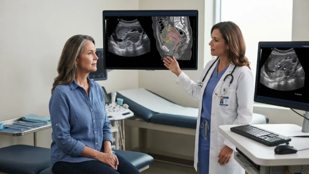
"A seemingly innocuous swallowed fish bone, lodging itself within the pelvic floor, can orchestrate a months-long deception, mimicking the insidious growth of a malignant tumor and presenting a formidable diagnostic challenge."
The human body, a marvel of intricate systems, can sometimes exhibit responses that confound even the most experienced medical professionals. In a rare and perplexing case reported in Frontiers in Oncology, a 55-year-old woman in Chengdu, China, experienced six months of intermittent, painless blood in her urine. This classic symptom, often a red flag for malignancy, led clinicians down a path of rigorous investigation for cancer. However, the true culprit was far more prosaic, yet equally dangerous: a fish bone, a seemingly small and common object, that had embarked on an extraordinary journey through her digestive and urinary tracts, culminating in a complex inflammatory mass that masterfully mimicked a deadly tumor. This case underscores the critical importance of considering an expansive differential diagnosis, even when symptoms strongly suggest a particular pathology, and highlights the diagnostic complexities that can arise from foreign bodies that migrate within the body.
The patient’s initial presentation was concerning. For half a year, she had noticed blood in her urine without any accompanying pain, a symptom she had unfortunately ignored. This period of asymptomatic hematuria was abruptly interrupted two days prior to her seeking medical attention. A sudden onset of heavy bleeding, coupled with increased urinary frequency and urgency, prompted her to finally consult physicians. An ultrasound revealed a significant abnormality: the posterior wall of her bladder was irregularly thickened, measuring approximately 5.3 by 1.9 by 5.5 centimeters. Crucially, the ultrasound detected dense blood flow signals within this thickened area, strongly suggesting a malignant tumor.
The medical team, operating under the assumption of cancer, proceeded with a transurethral resection biopsy. This procedure uncovered a broad-based, cauliflower-shaped growth, roughly 5 by 2 centimeters, on the bladder’s posterior wall. The mass was highly vascular and prone to bleeding, further reinforcing the initial diagnosis of a bladder tumor. However, the pathology report delivered an unexpected twist: the findings indicated chronic inflammation of the bladder lining with inflammatory granulation tissue, not cancerous cells. Further diagnostic tests, including white cell count, C-reactive protein levels, liver and kidney function tests, and tumor markers, all came back unremarkable. The patient had no prior surgical history that could explain the presence of a retained foreign object, and her cervical cytology was negative. Despite the biopsy’s findings, the bleeding returned three days later, leaving the medical team in a diagnostic quandary, as the situation no longer neatly fit either the initial suspicion of cancer or the biopsy’s findings of inflammation. This persistent and perplexing symptomology propelled the team to delve deeper with advanced imaging techniques.
The turning point in the diagnostic process was the persistent appearance of a thin, linear structure across multiple imaging modalities. A contrast-enhanced CT scan revealed the thickened bladder wall was seamlessly merging with the uterine isthmus, and between these structures, an unexplained linear dense shadow was observed. Subsequent MRI imaging depicted the same structure as a thin, dark line that did not absorb contrast. Radiologists interpreted this finding as either post-biopsy changes or, more intriguingly, a foreign body. A transvaginal ultrasound then provided further crucial detail, visualizing a hypoechoic mass, approximately 4.5 by 2.3 by 1.9 centimeters, occupying the space between the uterus and bladder. Within this mass, a bright, linear band was clearly visible, extending continuously from the uterine isthmus into the bladder wall.
The distinct appearances of this linear structure across different imaging techniques provided critical clues. It was bright on CT, dark on MRI, and bright on ultrasound. This characteristic pattern is indicative of a substance that is hard, dense, and acoustically resistant – qualities consistent with bone, a stone, or metal. Furthermore, the line’s continuous and regular nature argued against tumor calcification, which is typically more scattered. Instead, it pointed towards an object that did not belong in the body. The authors of the case report candidly admitted to a diagnostic trap: the initial imaging efforts were primarily focused on the interior of the bladder, where a tumor would typically be located, providing only limited views of the surrounding structures. The compelling clinical picture of painless hematuria had already steered the diagnostic pathway towards cancer, and the subsequent investigations naturally followed this established assumption.
The journey of a swallowed fish bone to the pelvic floor is an extraordinary and rare occurrence. Typically, ingested fish bones lodge in the throat or esophagus, causing immediate and severe discomfort. If they progress to the stomach, the acidic environment can often soften smaller fragments, allowing them to break down and pass through the digestive system. However, larger fragments retain their sharp edges. When such a fragment enters the lower digestive tract, the natural muscular contractions of the intestines (peristalsis) cannot break it down. Instead, peristalsis can drive the sharp object against the bowel wall, leading to ulceration and perforation. This perforation can result in serious complications such as peritonitis (inflammation of the abdominal lining), abscess formation, or the development of an inflammatory pseudotumor. Migrating bones have been documented to reach internal organs like the liver and pancreas, and in one prior report, a liver abscess caused by a fish bone was initially misdiagnosed as metastatic cancer.
What makes this patient’s case particularly unusual is the absence of typical symptoms associated with a perforated bowel. She experienced no throat discomfort, no abdominal tenderness, and no rebound pain, which are often indicative of peritonitis. Over several months, the bone acted as a persistent irritant, triggering a chronic inflammatory response in the surrounding tissues. This led to the formation of granulation tissue and scar tissue, which gradually enveloped the foreign object. This inflammatory encapsulation created a vascular, ill-defined mass that, on imaging, bore a striking resemblance to a tumor. While fish bones have been previously found embedded in the bladder wall, and one earlier report described a bone that perforated the rectosigmoid colon and subsequently migrated to the bladder during treatment, a single bone spanning and perforating three distinct organs – the rectum, uterus, and bladder – is considered exceptionally rare.
Surgical intervention was deemed necessary to remove the foreign body. The procedure was performed laparoscopically with endoscopic assistance. Upon exploration, the surgical team encountered a hard mass, approximately 5 by 3 centimeters, situated at the pelvic peritoneal reflection. The mass was severely inflamed and firmly adhered to the rectal wall, bladder, and uterine isthmus, obscuring the normal anatomical planes. Attempting to extract the bone intact, given its involvement across three organs, carried a significant risk of tearing and further damaging these vital structures. Therefore, the surgical strategy shifted. The surgeons meticulously separated the adhesions in stages, then precisely cut the bone into segments. These segments were subsequently removed through combined endoscopic access. The final pathological examination confirmed the presence of chronic inflammatory changes with no evidence of malignant cells.
Following the surgery, the patient’s recovery was rapid and complete. An ultrasound performed a week later showed only a scar-like area between the uterus and bladder. At one month post-operation, all her symptoms – the bleeding, urinary frequency, urgency, and painful urination – had resolved. A repeat CT scan of her abdomen and pelvis revealed no abnormalities. European guidelines for foreign body removal advocate for selecting endoscopic routes based on the patient’s symptoms, overall condition, and imaging findings, reserving open surgery for emergencies or cases of incomplete retrieval. A recent review of fish bone migration further elaborates on the diagnostic challenges associated with such cases.
While this case highlights the potential for foreign bodies to masquerade as malignancy, it is crucial to emphasize that unexplained blood in the urine is not typically caused by a foreign body. Painless hematuria remains a significant warning sign for bladder cancer and other serious urinary tract conditions, and it warrants prompt and thorough medical evaluation. The diagnostic journey in this particular case was a testament to the intricate interplay of clinical presentation, advanced imaging, and the often-unforeseen ways the body can react to foreign insults. The successful resolution underscores the importance of persistent clinical inquiry and the utilization of a multimodal diagnostic approach when faced with perplexing medical presentations.
Key Questions Answered
What was actually wrong with the patient?
A fish bone, approximately 3 centimeters in length, had perforated the patient’s rectum, the outer layer of her uterus, and her bladder wall, leading to the formation of a significant inflammatory mass.
Why was it mistaken for cancer?
The presence of painless blood in the urine, coupled with a highly vascular and ill-defined mass on the bladder wall observed through imaging, are classic indicators of bladder cancer. The chronic inflammation and granulation tissue that formed around the fish bone effectively mimicked the appearance of a tumor on medical scans.
What finally revealed it?
The breakthrough in diagnosis came with the consistent observation of a thin, linear structure across multiple imaging modalities. This line appeared dense on CT scans, dark on MRI, and bright on ultrasound, and it was traced continuously from the uterine isthmus into the bladder wall, strongly suggesting a foreign object.
How was it removed?
Surgeons employed a laparoscopic approach with endoscopic assistance. They carefully separated the adhesions binding the bone to the surrounding organs, then cut the bone into smaller segments. These fragments were then removed through combined endoscopic access, avoiding the risk of tearing the surrounding tissues that would have been involved in attempting to pull the bone out whole.
Did she recover?
Yes, the patient made a full recovery. One week after surgery, an ultrasound showed only a scar in the affected area. By one month post-operation, all her urinary symptoms had resolved, and a follow-up CT scan of her abdomen and pelvis was clear, showing no abnormalities.
Does this mean blood in the urine is often harmless?
No. Painless blood in the urine, known as hematuria, is a recognized warning sign for serious conditions, including cancer. It is essential that anyone experiencing unexplained blood in their urine seeks prompt medical evaluation to determine the underlying cause.
