
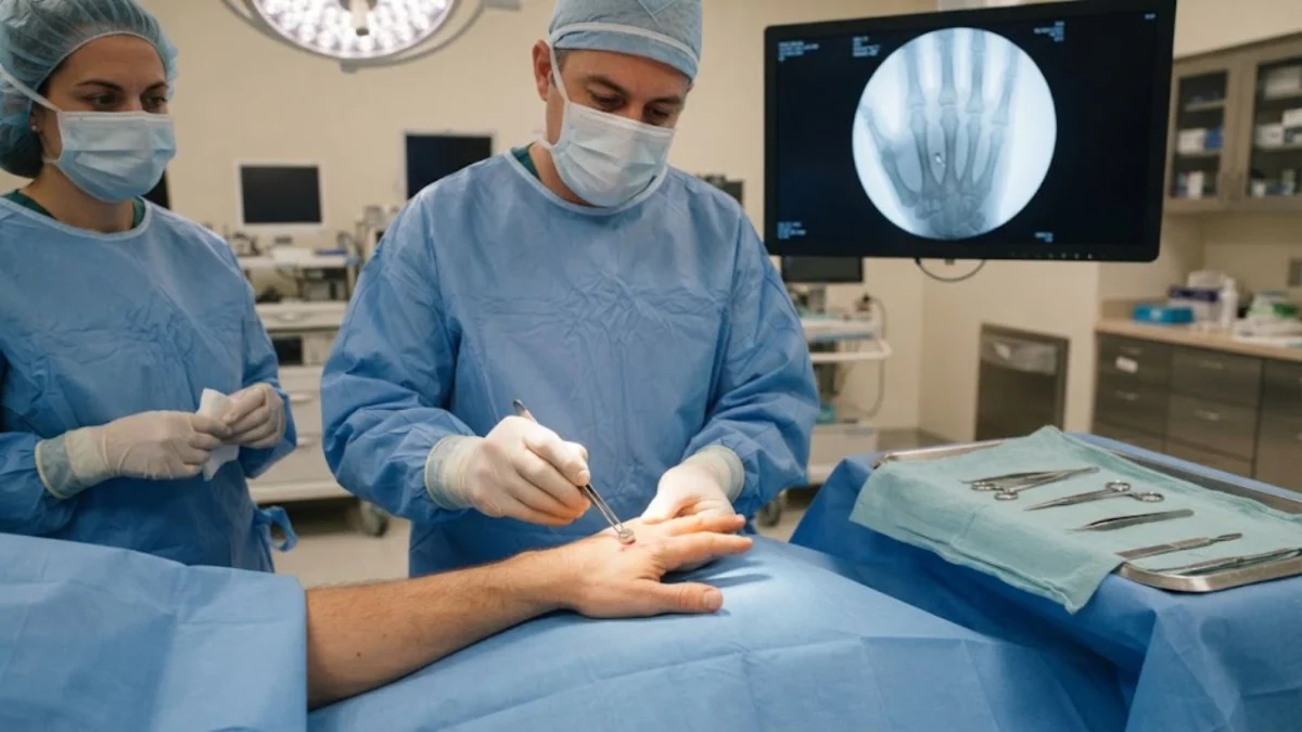
"Imagine a world where tiny metal shards lodged in your body could be coaxed out in minutes, with minimal radiation and often without a single X-ray. A new study reveals a surprisingly simple yet effective magnetic approach that could transform the treatment of common metallic foreign body injuries."
The challenge of removing embedded metal fragments from the human body has long been a complex medical undertaking. These fragments, often the result of industrial accidents, surgical mishaps, or even BB gun injuries, can lead to a cascade of complications including chronic pain, debilitating infections, and reactive scarring. Traditionally, extracting these foreign objects necessitates invasive surgery, involving meticulous dissection through soft tissues guided by multiple X-ray images, a process that is not only time-consuming but also carries its own inherent risks of damaging delicate nerves and blood vessels. However, a groundbreaking report from a team of surgeons in Istanbul is shedding light on a radically different, remarkably efficient, and significantly less invasive method: magnetic retrieval. This innovative technique leverages the power of magnets to locate and extract these troublesome metallic intruders, offering a glimpse into a future where complex procedures become streamlined and patient outcomes are significantly improved.
The core of this novel approach lies in the strategic application of sterilized, powerful magnets. Researchers have demonstrated that by carefully maneuvering a magnet over the skin’s surface, the precise location of a ferromagnetic fragment can be identified by the strength of its magnetic pull. Once pinpointed, a small incision is made, and the fragment is then drawn out directly to the magnet. This technique, detailed in a recent publication in Frontiers in Surgery, has been tested on 15 consecutive patients over a two-year period, yielding an astonishingly high success rate. Across this cohort, all 15 embedded fragments were successfully removed, with the average operative time clocking in at an impressive 3.13 minutes. Perhaps most remarkably, the average number of fluoroscopy images required per case was a mere 0.88, translating to a total of just 13 X-ray images for the entire series. This starkly contrasts with conventional methods that often demand numerous imaging sessions to guide the surgeon’s search.
The instruments employed in this magnetic retrieval method are surprisingly commonplace, readily available, and non-exotic. The primary tool is a commercially sourced neodymium-iron-boron disc magnet, measuring 10 millimeters in diameter and 1.5 millimeters in thickness, with a rating of N35 and a magnetic flux density between 1.17 and 1.20 tesla. For deeper or more dispersed fragments, the surgical team utilized a larger, 36-millimeter fishing magnet of the same grade. Crucially, both magnets were rigorously sterilized before each use to ensure patient safety and prevent infection.
The surgeons at the Istanbul hospital employed two distinct techniques, the choice of which was dictated by the specific characteristics of the wound and the fragment’s location. The first is termed "direct removal." In this scenario, the sterilized magnet is either placed directly into or beside a small incision, actively pulling the metallic fragment towards its surface. Patients and surgeons often report an audible or palpable "click" when the fragment makes contact with the magnet. When the fragment doesn’t immediately yield, the second technique, "vibration localization," comes into play. Here, the surgeon systematically sweeps the magnet across the affected area. The subtle shifting of the fragment within the tissue can be felt by the surgeon, allowing for a precise three-dimensional localization before forceps are introduced to complete the extraction. The decision on which technique to employ was made intraoperatively, based on a tactile assessment of the wound shape, the fragment’s depth, and the surgeon’s sensory feedback, rather than adhering to a rigid, pre-determined algorithm.
The vast majority of injuries treated with this magnetic approach were not dramatic in nature. Fourteen of the fifteen patients had sustained injuries from small ferromagnetic fragments, commonly encountered in metalworking environments, or from broken surgical needles that snapped during medical procedures. The hand was the most frequent site of these injuries, accounting for 12 out of the 15 cases. These hand procedures averaged a mere 2.75 minutes under local anesthesia, with a mean of just 0.6 fluoroscopy images. Cases involving the forearm and wrist, two in total, averaged slightly longer at 4.50 minutes. The single outlier was a more complex gunshot wound involving the forearm, wrist, and hand, which contained multiple spherical fragments. This case necessitated general anesthesia, required careful dissection around vital neurovascular structures, and involved four fluoroscopy images, extending the operative time to five minutes.
A significant advantage of this magnetic retrieval technique is its minimal reliance on anesthesia and imaging. Fourteen of the fifteen patients were treated under local anesthesia, a testament to the minimally invasive nature of the procedure. Twelve of these patients required only a superficial mini incision with no need for deep tissue dissection. Notably, no intraoperative complications were recorded during the study period. All patients were followed for a minimum of six months post-procedure, and all demonstrated excellent outcomes with no instances of infection, wound breakdown, or lingering nerve or vessel-related symptoms. The authors of the report emphasize that the drastically reduced number of fluoroscopy images is more than just an efficiency metric; it has profound implications for patient health, particularly for younger, working-age individuals who are disproportionately affected by these types of hand injuries. By minimizing cumulative radiation exposure, this technique contributes to a longer-term reduction in the potential risks associated with ionizing radiation.
However, it is crucial to acknowledge the limitations and specific prerequisites for the successful application of magnetic retrieval. The technique is exclusively effective for ferromagnetic metals; materials like aluminum, copper, and glass do not respond to magnetic forces. Therefore, a thorough understanding of the injury history and pre-operative imaging are essential to confirm the presence of an iron-based fragment before considering this approach. Furthermore, fragments that have been embedded in tissue for extended periods can develop a fibrous capsule around them, which can resist magnetic traction. In such cases, this capsule must be surgically opened before magnetic retrieval can be effective. Pre-operative confirmation of the fragment’s exact location through radiographs or ultrasound is not optional; it is a critical step in patient selection. The research team meticulously screened for these factors, accepting only patients whose fragments were visible on imaging and whose injury history suggested an iron-based metal. They also excluded individuals whose wounds required extensive cleaning due to contamination.
The geometry of the wound channel also plays a significant role. A fragment that entered the body at an oblique angle might be positioned in a location inaccessible through a straightforward incision, and forcing the approach could risk collateral damage. Within the delicate anatomical confines of the hand, the surgeons exercised particular caution, restricting the use of magnetic retrieval to superficial zones. They deliberately avoided areas in close proximity to critical structures such as digital nerve and artery bundles, flexor tendon sheaths, and small joint capsules. This precaution is due to the limited space available, which could lead to unpredictable acceleration of the fragment and potential injury to these vital components.
It is important to contextualize these findings appropriately. The study, while promising, involved a small series of 15 patients treated at a single hospital, utilizing retrospective records and without a direct comparison group. The authors themselves are forthright in describing their findings as "hypothesis-generating," acknowledging that definitive conclusions about whether this approach is universally faster, safer, or less radiation-intensive than conventional retrieval cannot be drawn without further comparative studies. Operative times are also influenced by factors such as surgeon experience and the complexity of individual cases, not solely by the technique itself.
The concept of using magnets for foreign body retrieval is not entirely new; it has roots dating back approximately a quarter-century, with initial descriptions appearing in the literature for metallic fragments in the neck. The technique has since been extended to various surgical specialties, including plastic surgery. The largest published experience to date, a substantial series encompassing 7,390 cases across mixed anatomical sites, reported an impressive 99.5 percent success rate. More recent research has continued to explore its utility, including a study of 22 adult limb cases with an average fragment depth of 2.35 centimeters and a pediatric extremity series that specifically highlighted the benefits of reduced imaging. What this new Istanbul-based paper uniquely contributes is the detailed breakdown of hand-specific operative times and imaging counts, extracted from a mixed-anatomy cohort for the first time. While the evidence is compelling, the authors and the broader medical community agree that prospective, comparative trials remain the crucial missing piece to firmly establish the superiority of magnetic retrieval in various clinical scenarios.
Key Questions Answered
What did the surgeons do?
Surgeons utilized sterilized rare earth magnets to precisely locate and extract embedded metal fragments from the hands, wrists, and forearms of 15 patients. The fragments were removed through small, minimally invasive incisions.
How fast was it?
The overall average procedure time, from the initial incision to wound closure, was an exceptionally brief 3.13 minutes. For hand-specific cases, the average duration under local anesthesia was even shorter, at 2.75 minutes.
Why does the number of X-ray images matter?
Conventional surgical retrieval of foreign bodies often requires repeated use of fluoroscopy (real-time X-rays) to guide the surgeon. This new technique significantly reduces radiation exposure, with the series averaging only 0.88 images per case. This is particularly beneficial for younger patients who have a longer lifetime exposure to consider.
When does the technique fail?
Magnetic retrieval is limited to ferromagnetic metals. It is ineffective for non-magnetic materials such as aluminum, copper, or glass. Additionally, fragments that have been lodged in tissue for a long time may become encased in a fibrous capsule, which must be surgically opened before magnetic extraction can be attempted.
Is this proven better than standard surgery?
Not definitively. The current study involved a small group of 15 patients and lacked a control group for direct comparison. The authors consider their findings to be hypothesis-generating, suggesting the need for larger, comparative studies to establish superiority in speed, safety, or radiation reduction.
Has this been done before?
Yes. The magnetic retrieval approach has been described and utilized for approximately 25 years. A large-scale study of 7,390 cases across various body parts previously reported a 99.5 percent success rate, indicating the technique’s established efficacy in broader applications.
