
"A lost IUD, initially missed by ultrasound, lay dormant within a woman’s bladder for nine years, leading to a rare case of migration and eventual surgical removal."
This remarkable medical case highlights the critical importance of accurate diagnostic imaging and thorough follow-up in reproductive healthcare. What began as a seemingly resolved abdominal discomfort a decade prior, turned out to be the initial, undetected migration of an intrauterine device (IUD) that had perforated the uterine wall and ultimately found its way into the bladder. The nine-year delay in diagnosis underscores the limitations of certain imaging techniques and the potential for rare but significant complications to go unnoticed, even in the absence of typical symptoms.
Unexplained Abdominal Pain and a Vanishing Device
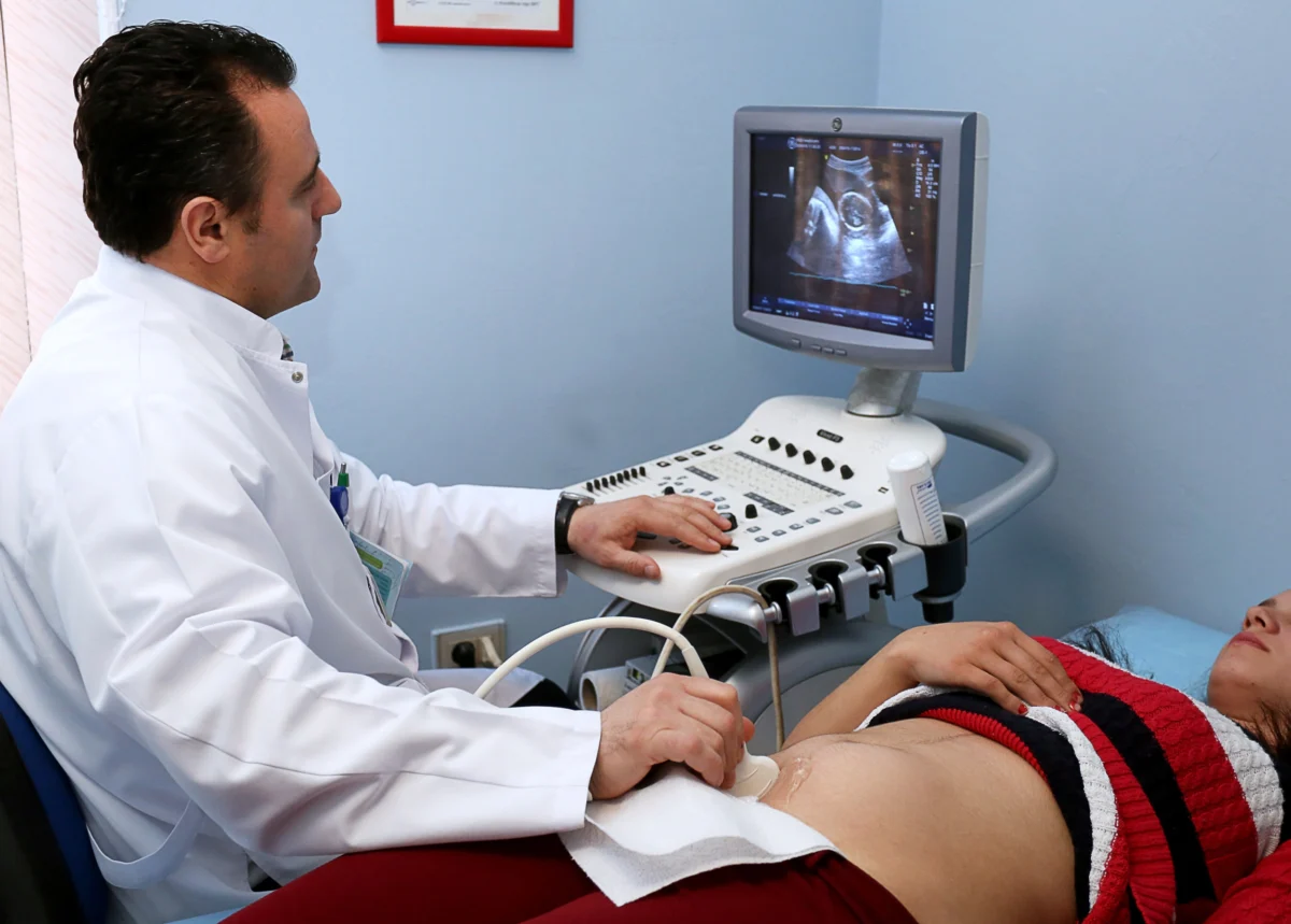
In March 2014, a woman in China received her fourth IUD insertion. Within a month, she presented with concerning symptoms: abdominal pain, swelling in her right leg, and difficulty with mobility. An initial ultrasound, the standard diagnostic tool for IUD placement, failed to locate the device within her uterus. Crucially, the ultrasound also revealed no abnormalities in the uterus or surrounding structures. After a period of rest, her symptoms subsided, and the case was seemingly closed, with the IUD’s whereabouts remaining a mystery.
This initial misinterpretation of the ultrasound, as later detailed by gynecologists from Shanghai University of Medicine and Health Sciences Affiliated Zhoupu Hospital and Gongshan People’s Hospital, proved to be a pivotal oversight. Their findings were published in Frontiers in Surgery on July 21, 2026, shedding light on this extraordinary case. The nine-year hiatus before the true nature of the problem was uncovered speaks volumes about the challenges of diagnosing subtle but serious medical conditions.
A History of Contraceptive Challenges and Unrecognized Pregnancies
The woman’s medical history, as outlined in the report, reveals a pattern of IUD expulsion. All four devices were inserted at the same local maternal and child health hospital. Prior to the problematic 2014 insertion, two IUDs placed in 2012 and a copper T in 2013 were each expelled within approximately two months. The fourth device, a copper IUD of the MYCu type, was inserted in March 2014. The report lists the timing and mechanism of its departure from the uterus as unknown.
Remarkably, in the nine years following this insertion, the woman experienced four more pregnancies. This included two full-term deliveries in 2017 and 2023, all while the IUD remained undetected outside of her uterus. This prolonged period of unrecognized uterine perforation and device migration highlights a significant gap in her subsequent medical care and the effectiveness of routine screenings in detecting such an anomaly.
Asymptomatic Migration: A Bladder Invader
When the woman was eventually admitted to the hospital at the age of 38, she presented with a week of persistent lower abdominal pain. Despite the IUD’s presence within her bladder, she reported no typical urinary symptoms such as frequency, urgency, pain, or blood in her urine. She also had no signs of infection like fever or chills. Her physical examination revealed only mild tenderness in the lower abdomen, and her blood work, including complete blood count and inflammatory markers, was within normal limits.
A diagnostic CT scan, employing three-dimensional reconstruction, was instrumental in revealing the true extent of the issue. It showed metallic densities within her bladder, with the largest fragment embedded in the bladder wall. Further investigation with a color Doppler ultrasound identified a distinct, bright structure measuring 15 by 6 millimeters inside the bladder.
Given that the hospital lacked laparoscopic equipment, surgeons opted for an open abdominal procedure. During surgery, they discovered dense adhesions binding the omentum, a fatty tissue apron in the abdomen, to the bladder. The IUD was found to be intact. One arm of the device was lodged within the muscular layer of the bladder wall, while the other arm had fully penetrated the wall, extending into the bladder’s interior. The surgical team successfully removed the IUD and closed the bladder in two layers. She was discharged five days later, and a follow-up ultrasound in February 2024 confirmed a normal bladder. The authors of the report noted that she has remained symptom-free.
Understanding IUD Perforation: Causes and Consequences
The authors of the case report propose that the most plausible explanation for the IUD’s migration is a "procedural error during insertion." This suggests that the device may have been improperly placed, leading to perforation of the uterine wall at the time of insertion. It is important to note that the woman had no history of uterine perforation prior to this event, and her insertions did not occur during the postpartum period while breastfeeding, a time that has been associated with a slightly increased risk of perforation.
Uterine perforation by an IUD, while a known complication, is relatively uncommon. A comprehensive European study involving over 61,000 IUD users reported perforation rates of 1.4 per 1,000 insertions for hormonal devices and 1.1 per 1,000 insertions for copper devices. While breastfeeding at the time of insertion did elevate the relative risk approximately sixfold, the absolute risk remained low, and importantly, no perforations in that study resulted in severe illness or organ injury.
The limitations of ultrasound in detecting displaced IUDs are also highlighted by this case. While ultrasound is the primary imaging modality for IUD placement and follow-up, it can sometimes fail to accurately pinpoint a migrated device. The authors reviewed published cases where color Doppler ultrasound exhibited missed-diagnosis rates as high as 28% for displaced devices, whereas CT scans consistently located them. In two earlier reported cases of IUDs found in the bladder, Doppler ultrasound had failed to detect devices that CT scans subsequently identified years after insertion. This underscores the critical point that an empty uterus on ultrasound does not definitively mean the IUD is absent.
A review of 30 published studies on IUDs found in the bladder revealed that urinary tract infections and lower abdominal or pelvic pain were the most frequently reported symptoms, typically leading to earlier diagnosis. However, a subset of patients, like the one in this case, presented with no symptoms. The time to displacement varied significantly, ranging from 1 to 312 months after insertion, with reported peaks occurring within the first year and again after five years. Copper T and MYCu devices were the types most frequently implicated, which the authors attribute to their widespread use and the presence of rigid side arms that may exert pressure on the uterine wall. The research was supported by public science and health programs in Shanghai’s Pudong New Area, with the authors declaring no commercial conflicts of interest.
Lessons Learned for IUD Users and Healthcare Providers
Intrauterine devices remain a highly effective and widely utilized form of long-acting reversible contraception. Complications like the one described are exceptionally rare. This report details a singular case involving a specific device type and medical care provided in a setting lacking advanced laparoscopic equipment, thus it cannot definitively quantify the incidence of such events in other regions, including the United States.
However, this case reinforces a crucial message echoed in other medical reports. Doctors affiliated with Aga Khan University previously described a 40-year-old woman whose copper IUD was presumed to have been expelled after it was not visible on ultrasound. Approximately a year later, she received a hormonal IUD. Subsequent imaging revealed that the initial device had migrated outside the uterus, necessitating abdominal surgery for its retrieval. This experience led the authors to strongly advocate for confirmatory imaging whenever an IUD cannot be visualized.
This approach is increasingly integrated into clinical guidelines. A July 2026 algorithm developed by the Reproductive Health Access Project for managing IUDs with unseen strings begins with an ultrasound. If the device is not visible, an abdominal X-ray is recommended. If the IUD is confirmed to be outside the uterus, patients are referred for surgical intervention.
For individuals using IUDs, the practical advice is clear and actionable. Anyone who cannot locate their IUD strings or who experiences new pelvic pain, urinary symptoms, or other unexplained health issues following an IUD insertion should promptly consult the clinician who placed the device. Diagnostic imaging can accurately determine the IUD’s location and address any potential complications. This case serves as a stark reminder that while IUDs are generally safe and effective, vigilance and comprehensive diagnostic evaluation are paramount in ensuring patient well-being.
