
"A seemingly straightforward case of abdominal pain in a young girl highlights the diagnostic challenges posed by rare conditions, where subtle imaging clues can mislead even experienced physicians towards more common, yet ultimately incorrect, diagnoses."
A seven-year-old girl’s persistent, intermittent upper abdominal pain, coupled with a profound sense of fullness after minimal food intake and a significant weight loss of nearly 10 pounds, led her doctors down a diagnostic path that initially pointed towards a rare vascular condition. However, the true culprit, a remarkably large hairball measuring almost four inches, was eventually uncovered, underscoring the complexities of pediatric gastrointestinal disorders and the importance of comprehensive diagnostic approaches. This compelling case, detailed in a recent publication in Frontiers in Pediatrics by medical professionals from The Valley Hospital and Valley Medical Group in Paramus, New Jersey, serves as a crucial reminder that even seemingly unusual conditions like gastric trichobezoars can meticulously mimic more prevalent syndromes, particularly on advanced imaging studies.
The previously healthy child presented with a constellation of symptoms that, while concerning, lacked definitive markers for immediate diagnosis. Her diminished appetite was notable, though she maintained adequate fluid intake, ruling out dehydration as a primary cause. Crucially, she exhibited no signs of vomiting or diarrhea, which often accompany more acute gastrointestinal blockages. This absence of typical obstructive symptoms added another layer of complexity to the diagnostic puzzle.
Initial investigations, including blood work, revealed subtle anomalies. A mildly elevated platelet count can indicate a response to inflammation or stress. A slightly elevated lipase level, an enzyme crucial for fat digestion, could suggest pancreatic irritation or other gastrointestinal distress. The most striking finding was iron deficiency, evidenced by an iron saturation of a mere 5%, pointing to a potential source of chronic blood loss or malabsorption. Despite these findings, her vital signs remained stable, and she was afebrile, with no fever. A routine abdominal X-ray and an ultrasound of the abdomen yielded normal results, offering no immediate clues to the underlying cause of her discomfort.
Given the persistence of her symptoms and the concerning weight loss, her medical team escalated their diagnostic efforts by ordering an Magnetic Resonance Imaging (MRI) scan. This decision was strategic, aiming to gather detailed anatomical information without exposing the child to the radiation associated with a Computed Tomography (CT) scan. The MRI revealed significant dilation of the stomach and the duodenum, the initial segment of the small intestine. More importantly, it showed what appeared to be a narrowing at the point where the superior mesenteric artery (SMA) crosses the duodenum. These findings were interpreted as highly suggestive of Superior Mesenteric Artery (SMA) syndrome.
SMA syndrome is a relatively rare condition characterized by compression of the third part of the duodenum by the superior mesenteric artery. This compression can occur when the normal cushioning fat pad surrounding the artery diminishes, often following rapid weight loss. The syndrome predominantly affects young females and shares several clinical manifestations with trichobezoars, including nausea, vomiting, upper abdominal pain, and a feeling of premature fullness after eating. This overlap in symptoms and imaging findings contributed significantly to the initial misdiagnosis.
Further compounding the diagnostic uncertainty, an upper gastrointestinal X-ray series, which involves ingesting a barium contrast agent, reinforced the suspicion of SMA syndrome. This study demonstrated delayed gastric emptying and a dilated duodenum, with some improvement noted when the patient’s position was altered. These observations are characteristic of mechanical obstruction in the duodenal region.
However, one persistent anomaly in the imaging studies began to sow seeds of doubt. The X-ray series consistently showed a "filling defect" within the stomach, a region that appeared to be occupied by something other than stomach contents or air. This finding, coupled with the discrepancy between the severity of the patient’s symptoms and the inferred severity of SMA syndrome, prompted the medical team to consider an alternative diagnostic approach. Recognizing that imaging alone was not providing a definitive answer, they proceeded with an upper endoscopy.
The upper endoscopy, a procedure that involves inserting a flexible tube with a camera into the esophagus, stomach, and duodenum, proved to be the pivotal diagnostic step. Visual examination revealed the true nature of the problem: a large trichobezoar, a mass composed of ingested hair, lodged within the main body of the stomach. The endoscopy also identified significant ulceration surrounding the hairball, which provided a direct explanation for the patient’s iron deficiency anemia. The authors of the report highlighted that the ulceration was likely caused by the constant friction and pressure exerted by the large hair mass on the stomach lining.
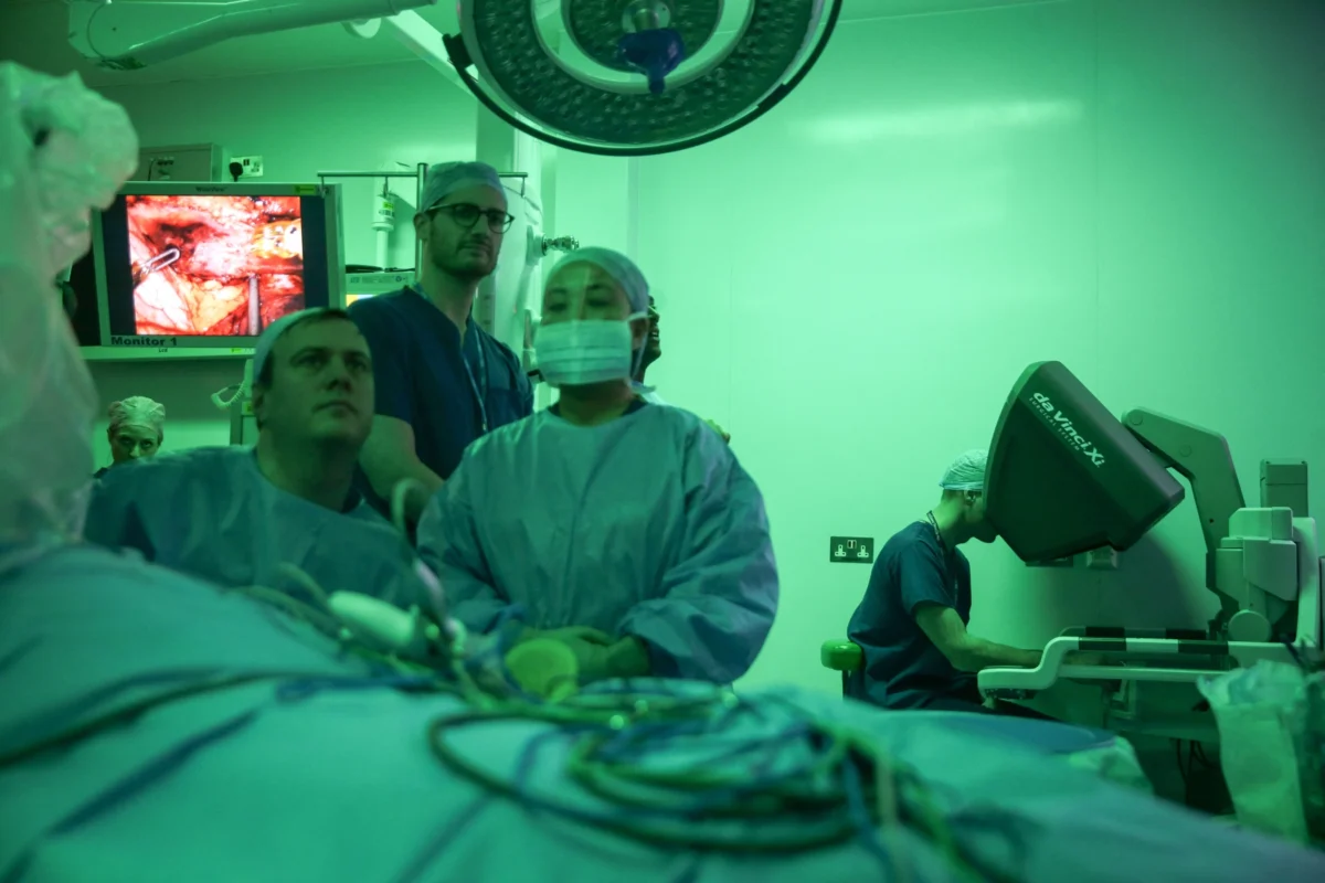
Due to the considerable size and density of the trichobezoar, attempts to remove it endoscopically were deemed unsafe and impractical. Consequently, the surgical team opted for a laparoscopic-assisted procedure. This minimally invasive technique involved making small incisions in the abdomen through which surgical instruments and a camera could be inserted. The stomach was then opened, and the formidable hairball was carefully extracted in a single piece. Its dimensions were measured at an impressive 10.0 by 3.2 by 3.0 centimeters, a significant mass for a child of this age.
The surgical intervention was successful, and the patient’s recovery was uneventful. She was able to resume oral intake and was prescribed an acid-reducing medication to aid in the healing of the stomach ulcers. At her one-week follow-up appointment, she was reportedly eating well, had already regained approximately five pounds, and was completely free of abdominal pain.
The underlying cause of the trichobezoar was subsequently elucidated through discussions with the child’s parents. They revealed that the girl had a habit of sucking on and swallowing her hair, particularly during periods of anxiety. This behavior had not been readily apparent to the parents, and the absence of overt hair loss or a pre-existing psychiatric diagnosis had initially masked the behavioral component of her illness. The authors emphasize that the lack of initial suspicion was understandable, as such habits can be subtle and are not always accompanied by visible signs.
To address the root cause and prevent recurrence, the child was referred for outpatient behavioral health counseling. Her parents received guidance on monitoring her behavior and strategies to mitigate the risk of future hair ingestion. Thankfully, at the one-week follow-up, no further instances of hair-eating were reported. The authors underscore the critical importance of a multidisciplinary approach involving pediatric surgery, gastroenterology, and behavioral health specialists to ensure long-term positive outcomes and to address the underlying behavioral patterns that contribute to trichobezoar formation.
The diagnostic challenges posed by trichobezoars are further illuminated by their relative rarity and the limitations of standard imaging techniques. Trichobezoars occur in less than 1% of children, with a predilection for young girls. Plain X-rays have a low sensitivity for detecting these masses, identifying them in only 10% to 18% of cases. Ultrasound can also be hampered by trapped air and food particles within the stomach, obscuring visualization.
While contrast-enhanced CT scans are considered the preferred imaging modality for bezoars, offering a sensitivity of up to 90%, their use in young children is often weighed against concerns regarding radiation exposure. In this specific case, the decision to defer CT in favor of MRI was driven by these radiation concerns. Although MRI avoids radiation, its accuracy in detecting bezoars is less established. Large hairballs can appear as dark, amorphous areas on MRI, easily mistaken for air or other non-specific findings within the stomach, as was the case here where the MRI did not clearly depict a distinct mass.
In retrospect, the medical team acknowledges that their initial diagnosis of SMA syndrome was based on the confluence of symptoms and imaging features, rather than definitive proof of arterial compression. The fundamental difference between SMA syndrome and a trichobezoar lies in the mechanism of obstruction: SMA syndrome involves external compression of the duodenum, whereas a trichobezoar creates an internal blockage. Endoscopy, therefore, remains the definitive diagnostic tool for trichobezoars.
The potential complications of undiagnosed trichobezoars can be severe. If left untreated, these hair masses can extend from the stomach into the small intestine, a condition known as Rapunzel syndrome. This rare variant significantly increases the risk of serious complications, including ulceration, bleeding, damage to the intestinal wall, and even perforation, a life-threatening emergency. A case report and literature review published in the same journal highlights Rapunzel syndrome as an exceedingly rare cause of digestive distress in children.
While this single case report cannot quantify the frequency with which trichobezoars are mistaken for SMA syndrome, it serves as a compelling illustration of the diagnostic quandaries that can arise. Parents who observe their children engaging in hair-chewing or swallowing behaviors, particularly when accompanied by symptoms such as abdominal pain, early satiety, poor appetite, or unexplained weight loss, are strongly encouraged to raise these concerns with their pediatrician. Early recognition and intervention are paramount in ensuring timely diagnosis and appropriate management.
What is a trichobezoar?
A trichobezoar is a concretion or mass formed from ingested hair that accumulates in the stomach. Hair is resistant to digestion, and the stomach’s normal muscular contractions are often insufficient to break down and propel such masses through the digestive tract.
Why did doctors first suspect SMA syndrome?
The initial suspicion of SMA syndrome stemmed from the MRI and upper gastrointestinal X-ray series, which showed a distended stomach and duodenum with apparent narrowing at the point where the superior mesenteric artery crosses the duodenum. These imaging findings are characteristic of SMA syndrome, a vascular compression of the small intestine.
How was the hairball found and removed?
An upper endoscopy, a procedure allowing direct visualization of the upper digestive tract, revealed the presence of a large hairball within the stomach. Surgical intervention was then performed using a laparoscopic-assisted approach, where the stomach was opened to allow for the removal of the hairball in one piece.
What warning signs should parents know?
Parents should be vigilant for signs of hair chewing or swallowing in their children, especially when these behaviors are accompanied by symptoms such as persistent stomach pain, a feeling of fullness after eating very little, poor appetite, or unexplained weight loss. Such a combination of factors warrants prompt consultation with a pediatrician.
Can hairballs come back?
The risk of recurrence exists if the underlying behavioral habit of hair-eating is not addressed. The girl in this case was referred for behavioral health counseling to manage this underlying issue and reduce the likelihood of future trichobezoar formation.
