
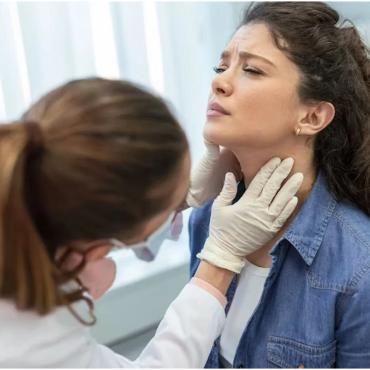
"A lump near the jaw, while concerning, is often misdiagnosed as a more serious condition like a brain tumor, highlighting the critical importance of precise medical terminology and understanding distinct anatomical structures."
The recent public clarification by Dez Fafara, frontman of the bands DevilDriver and Coal Chamber, regarding a non-cancerous tumor on his salivary gland serves as a stark reminder of how easily medical findings can be misinterpreted, leading to unnecessary alarm. Fafara took to social media to correct reports that erroneously stated he had a brain tumor, emphasizing that his condition involves a tumor "THAT’S NOT CANCER ON MY SALIVA GLAND." This distinction is far from a mere technicality; it represents fundamentally different medical scenarios involving distinct organs, specialized medical fields, varying prognoses, and different surgical approaches. The understandable confusion arises from the fact that Fafara’s tumor was discovered during an MRI ordered after a vertigo episode, and the potential surgical risks he described—such as impacts on blinking, smiling, and speaking—sound alarmingly neurological, mirroring concerns associated with brain surgery. However, these neurological implications stem from the intricate relationship between the salivary gland and the facial nerve, a crucial anatomical connection that dictates the management and potential complications of parotid gland tumors.
The anatomy of the parotid gland is central to understanding why tumors within it can present with symptoms that evoke concerns typically associated with neurological conditions. The parotid glands, the largest of the salivary glands, are situated just in front of and below each ear, extending towards the jawline. Crucially, the facial nerve, also known as the seventh cranial nerve, emerges from the base of the skull and traverses directly through the parotid gland before branching out to innervate the muscles of facial expression. These branches are responsible for a myriad of movements, including raising the eyebrows, closing the eyelids, moving the corners of the mouth, and controlling the lower lip. Damage to any of these branches can result in weakness or paralysis in the corresponding facial area. The eyelid branch is of particular significance due to the vital role of a fully closing eyelid in protecting the cornea from injury.
This anatomical proximity explains why Fafara’s description of potential surgical risks—affecting "my blinking, my smiling, my speaking"—aligns with neurological concerns. A surgical procedure to remove a parotid gland tumor is, in essence, a delicate exercise in nerve dissection. The surgeon’s primary objective is to meticulously identify the main trunk of the facial nerve and meticulously trace each of its branches, carefully separating the tumor tissue from the delicate nerve fibers. While temporary facial weakness is a common post-operative occurrence, permanent weakness, though less frequent, is a recognized risk, particularly when the tumor is large, deeply situated within the gland, or if it is a recurrence requiring a second surgical intervention.
Salivary gland tumors, in general, are relatively uncommon, and their likelihood of being malignant varies significantly depending on the specific gland involved. Within the parotid gland, the vast majority of tumors are benign. The most frequently diagnosed benign parotid tumor is pleomorphic adenoma. However, as the size of the salivary glands decreases, the proportion of malignant tumors increases. Tumors affecting the submandibular glands, located beneath the jaw, are malignant more often than those in the parotid glands. The highest malignancy rates are associated with tumors of the minor salivary glands, which are scattered throughout the oral cavity and throat.
The diagnostic pathway for salivary gland masses typically involves imaging studies, with magnetic resonance imaging (MRI) being particularly valuable for its detailed visualization of soft tissues. This is often complemented by obtaining a tissue sample through fine needle aspiration (FNA) or a core biopsy. This combination of imaging and tissue analysis is essential for distinguishing between benign and malignant tumors and forms the basis for all subsequent treatment decisions. Fafara has undergone such testing and has received a non-cancerous diagnosis.
Several clinical features can raise suspicion for malignancy. These include rapid tumor growth, the presence of pain, fixation of the mass to surrounding tissues, skin involvement, enlarged lymph nodes in the neck, and, most critically, pre-existing facial weakness or numbness. A tumor that is already impairing nerve function suggests a more aggressive behavior, which is atypical for most benign salivary gland masses.
In select patient populations, watchful waiting, or observation, is a legitimate management strategy for benign salivary gland tumors. This approach is generally considered for older patients, individuals with significant co-existing medical conditions that increase surgical risk, cases involving small tumors in anatomically challenging locations, or when patients, after thorough consultation and understanding of the potential trade-offs, opt for observation. The rationale behind observation is that the surgery itself carries the inherent risk of facial nerve damage, and in some patients, this risk may outweigh the benefits of removing a slow-growing benign mass.
The decision to observe or operate involves careful consideration of the trade-offs. Pleomorphic adenomas, for instance, tend to grow slowly but have the potential for malignant transformation over extended periods. Therefore, observation necessitates regular interval imaging to monitor for any changes. Larger tumors can also present greater surgical challenges, potentially increasing operative difficulty and risks. Fafara’s statement that his doctors advised him that the tumor needs to grow before it can be safely removed is an unusual rationale, as surgeons more commonly cite tumor size as a reason to proceed with surgery sooner rather than later to minimize potential complications. It is crucial for individuals to understand that the specific location, depth, and relationship of a salivary tumor to the facial nerve can significantly alter the surgical calculus, making any generalized advice inapplicable to individual cases.
Furthermore, Fafara’s mention of working to shrink the tumor through diet, positivity, and prayer, and reporting that it has been shrinking, warrants careful contextualization. While maintaining a positive outlook and engaging in self-care practices can be profoundly beneficial for overall well-being, no dietary interventions have been scientifically proven to shrink salivary gland tumors. Apparent changes in tumor size between imaging scans can often be attributed to variations in measurement techniques, imaging protocols, or natural fluctuations in tumor volume. The psychological and emotional benefits derived from faith and proactive engagement in one’s health are undeniable and valuable; however, these should not be misconstrued as direct tumor treatments, nor should they lead to the delay of professional medical evaluation for any persistent neck or jaw mass.
Any new lump detected in front of or below the ear, along the jawline, or under the jaw that does not resolve within two to three weeks warrants prompt medical evaluation by a clinician. While most such lumps are benign and may represent swollen lymph nodes or blocked salivary ducts, persistent lumps necessitate investigation. Features that indicate a need for more urgent attention include a rapidly growing mass, pain in the affected area, any facial numbness or weakness, a lump that feels fixed rather than mobile, difficulty opening the mouth, or a lump accompanied by swollen lymph nodes in the neck.
The appropriate specialist for evaluating a suspected salivary gland tumor is an otolaryngologist, commonly known as an ear, nose, and throat (ENT) surgeon, and specifically a head and neck surgeon for tumors. A primary care physician can provide a referral to the appropriate specialist. Imaging and biopsy procedures typically require a referral, making this the practical initial step in the diagnostic process.
Regarding the financial aspects of diagnosis, MRI scans and biopsies for documented neck masses are generally covered by commercial health insurance and Medicare when deemed clinically indicated. However, prior authorization for MRI is a common requirement. Patients facing insurance denials can often secure approval by having their ordering clinician document the specific physical findings and the duration of symptoms, which are key factors in determining medical necessity.
In Fafara’s case, the specific type of tumor has not been publicly disclosed, and his account of the tumor shrinking lacks accompanying clinical documentation. While he intends to continue touring, MedicalDaily, like other reputable health news outlets, does not report on individuals’ private medical care beyond what they voluntarily disclose. The distinction between a benign salivary gland tumor and a malignant brain tumor is crucial, impacting prognosis, treatment, and overall patient outcomes. Understanding the distinct anatomy and pathology of these conditions is paramount for accurate diagnosis and effective management.
